|
|
| Contents | Post-Test |
By definition, regeneration of the periodontium requires the following:21
For more than a decade, periodontal research has been studying the potential of growth factors and bone morphogenetic proteins (BMPs) to achieve predictable periodontal regeneration, yet the work is still in the early stages of development. Bone morphogenetic proteins have demonstrated distinctive properties that contribute to the formation of new cementum and ectopic bone formation.21
Research has produced a much greater collection of published clinical and histologic data for animal studies than for human studies. In 1991, osteogenin (bone morphogenic protein-3) in combination with DFDBA (demineralized freeze-dried bone allograft) was used in the first human trials. The results demonstrated that, in a submerged environment, this combination was able to significantly enhance the regeneration of a new attachment.21
The results from the human trials were substantiated by various animal studies that also revealed increased regeneration when bone morphogenetic proteins-2 and -7 were utilized to treat periodontal defects. However, in animal studies, a higher incidence of ankylosis was reported. One study noted that 15 out of 17 dogs developed ankylosis after treatment with bone morphogenetic protein-2. Yet, when sites were treated with bone morphogenetic protein-7, investigators did not observe ankylosis. Further human clinical and histologic studies are recommended to better evaluate the potential of these agents to induce periodontal regeneration.21
There are other growth factors that primarily act as a mitogen or differential factor on regenerating periodontal tissues, including: 21
Human clinical studies were conducted to evaluate the use of recombinant platelet derived growth factor and insulin-like growth factor. The published data reported mixed results when these growth factors were used to treat periodontal intraosseous defects or furcations. Treatment with recombinant platelet derived growth factor and insulin-like growth factor achieved the best results in furcations, producing approximately 42% bone fill nine months after surgery.21
The system used to deliver growth factors may have a significant impact on the regenerative response that is achieved. Investigators are especially interested in the surface area, surface properties for cell-surface interactions, inflammatory and immune reactions, and degradation kinetics. Delivery systems that have been reported are collagen as a sponge, membrane, or gel and gelatin with varying degrees of cross-linking.21
The fact that bone and cementum formation take place over different time periods in animal models must be allowed for during drug delivery. The degradation kinetics of bioabsorbable carriers, or how fast they break down, appear to affect or modify the type of new tissue that is formed. For example, bone formation was induced to a greater extent by a fast degradation and fast release of bone morphogenetic protein-2. In contrast, the formation of cementum increased significantly in the presence of the slow degrading and slow releasing bone morphogenetic protein gelatin carrier.
In view of the inadequate amount of human clinical data that is presently available, scientists have yet to determine if these findings are applicable to humans in the environment of inflammation associated with periodontal disease.21
The reduction or elimination of tissue inflammation induced by bacterial plaque and its by-products, correction of defects or anatomical problems caused by the disease process, and regeneration of lost periodontal tissues as a consequence of disease destruction are all important goals of periodontal therapy. The process of periodontal regeneration is certainly complicated by factors such as the presence of multiple, specialized cell types and attachment complexes, stromal-cellular interactions, diverse microbial flora, and avascular tooth surfaces.21
Although only limited human clinical data and limited long-term follow- up are available, growth factors and proteins have shown promising results in pre-clinical trials. Before this treatment can be recommended as a routine clinical procedure, additional studies are needed to establish clinical efficacy and long-term stability.21
Efforts continue in the direction of enhanced understanding of periodontal regeneration biology, and scientists anticipate that developments in biologic and materials sciences will result in new guided tissue regenerative materials and delivery systems. The ultimate success of regenerative therapies is dependent upon establishing a scientifically sound, evidence-based rationale, as well as adequate human clinical data.21
Apoptosis - a cell biology term that means “programmed cell death as signaled by the nuclei in normally functioning human and animal cells when age or state of cell health and condition dictates. It is an active process requiring metabolic activity by the dying cell, often characterized by cleavage of the DNA into fragments that give a so called laddering pattern on gels. Cells that die by apoptosis do not usually elicit the inflammatory responses that are associated with necrosis, though the reasons are not clear”.3
BSP (bone sialoprotein) and OPN (osteopontin) - “The two major non-collagenous proteins found in cementum are bone sialoprotein (BSP) and osteopontin (OPN). These proteins remain bound to the collagen matrix, and they demonstrate cell attachment properties via the arg-gly-asp (RGD) sequence. Bone sialoprotein and osteopontin are prominently expressed in acellular extrinsic fiber cementum and acellular afibrillar cementum. Cells located along the root surface express both of these proteins during early tooth root development. Bone sialoprotein is expressed by root surface cells and it is also found in mature teeth. Osteopontin is found within the periodontal ligament area that is associated with mature teeth.”11
CAP - “cementum attachment protein (CAP) which is a collagenous protein, unique to cementum. Expressed by cementoblasts in bovine tooth germs, cementum attachment protein stimulates the adhesion and spreading of mesenchymal cells, however, it preferentially stimulates the adhesion of mineralized-tissue-forming cells.”11
cDNA - “an acronym for complementary DNA that is synthesized from a messenger RNA template, the single-stranded form is often used as a probe in physical mapping to locate the gene or can be cloned in the double stranded form. Viral reverse transcriptase can be used to synthesize DNA that is complementary to RNA (for example an isolated mRNA).”3
Constitutively expressed - means “constantly present, whether there is demand or not, as opposed to inducible.”3
Downstream - “a molecular biology term indicating portions of DNA or RNA that are more remote from the initiation sites and that will therefore be translated or transcribed later. It is a shorthand term for things that happen at a late stage in a sequence of reactions.3 The use of the term downstream is a convention used to describe features of a DNA sequence, gene or mRNA related to the position and direction (5 to 3) of transcription by RNA polymerase or translation by the ribosome. Downstream (or 3 to) is in the direction of transcription (or translation) whereas upstream (5 to) is in the direction from which the polymerase (or ribosome) has come. Conventionally DNA sequences, gene maps and RNA sequences are drawn with transcription (or translation) from left to right and so downstream is towards the right.”20
Explant - “a living tissue that has been transferred from an organism to an artificial medium for culture.”3
Ex vivo - in vitro
Fibronectin - “a protein, specifically a high molecular weight glycoprotein. It occurs in an insoluble fibrillar form in extracellular matrix of animal tissues and a soluble form in plasma. Because of their multiple domains, fibronectins have the ability to interact with a variety of extracellular substances such as collagen, fibrin and heparin and with specific membrane receptors on responsive cells. Noteworthy are the RGD domain that is recognized by integrins and two repeats of the EGF like (epidermal growth factor like) domain. When a cell’s fibronectin receptors (members of the integrin family) interact with fibronectin adsorbed to a surface, the result is adhesion and spreading of the cell.”3
Gla - “4-carboxyglutamic acid. A carboxylated form of glutamic acid found in certain proteins (e.g., prothrombin, factors VII, IX, and X, osteocalcin). Its synthesis is vitamin K-dependent.”3
Heparan sulfate (HS) - “a linear polysaccharide found in all animal tissues. It occurs as a proteoglycan (PG) in which two or three HS chains are attached in close proximity to cell surface or extracellular matrix proteins. It is in this form that HS binds to a variety of protein ligands and regulates a wide variety of biological activities, including developmental processes, angiogenesis, blood coagulation and tumour metastasis.”19
Heparan sulfate proteoglycan - “Ubiquitous macromolecules associated with the cell surface and extracellular matrix of a wide range of cells of vertebrate and invertebrate tissues. They are essential cofactors in cell-matrix adhesion processes, in cell-cell recognition systems, and in receptor-growth factor interactions.”3
Heparin - “Sulphated mucopolysaccharide, found in granules of mast cells, that inhibits the action of thrombin on fibrinogen by potentiating antithrombins, thereby interfering with the blood clotting cascade. Platelet factor IV will neutralize heparin.”3
In situ - a term that means “in the natural or normal place, confined to the site of origin without invasion of neighboring tissues”.3
Isoform - a protein that has “the same function and similar (or identical sequence), but is the product of a different gene and is usually tissue specific. The term isoform carries a stronger implication than the term homologous”.3
Knockout - an “informal term for the generation of a mutant organism in which the function of a particular gene has been completely eliminated (a null allele)”.3
Lipopolysaccharide – “a major constituent of the cell walls of gram-negative bacteria”3, i.e., periodontal pathogenic bacteria; it is associated with loss of alveolar bone in periodontal disease. “Lipopolysaccharide inhibits the proliferation of gingival fibroblasts; and platelet-derived growth factor decreases this inhibitory effect.”1
Matrix metalloproteinases (MMPs) - “a family of nine or more highly homologous Zn(++)-endopeptidases that collectively cleave most if not all of the constituents of the extracellular matrix.”25
Mechanotransduction - “refers to the many mechanisms by which cells convert mechanical stimulus into chemical activity.”19
Mitogen - “a substance capable of inducing mitosis of certain eukaryotic cells”3
OCN - osteocalcin - “Polypeptide of 50 residues formed from a 76-77 amino acid precursor and found in the extracellular matrix of bone. Binds hydroxyapatite.3 Osteocalcin is a noncollagenous protein found in bone and dentin. It is secreted by osteoblasts and thought to play a role in mineralization and calcium ion homeostasis. It has been stipulated that osteocalcin may also function as a negative regulator of bone formation, although its exact role is unknown.”19
OPN - osteopontin - see BSP above.
Orthotopic - “in the normal or usual position.”3
Osteoconduction – “means that bone grows on a surface. This phenomenon is regularly seen in the case of bone implants. Implant materials of low biocompatibility such as copper, silver and bone cement shows little or no osteoconduction. Osseointegration is the stable anchorage of an implant achieved by direct bone-to-implant contact. In craniofacial implantology, this mode of anchorage is the only one for which high success rates have been reported.”24
Osteoinduction - “is the process by which osteogenesis is induced. It is a phenomenon regularly seen in any type of bone healing process. Osteoinduction implies the recruitment of immature cells and the stimulation of these cells to develop into preosteoblasts. In a bone healing situation such as a fracture, the majority of bone healing is dependent on osteoinduction.”24
Osseointegration - “is the growth action of bone tissue, as it assimilates surgically in implanted devices or prostheses to be used as either replacement parts (e.g., hip) or as anchors (e.g., endosseous dental implants).3 Osseointegration is possible in other parts of the body, but its importance for the anchorage of major arthroplasties is under debate. Ingrowth of bone in a porous-coated prosthesis may or may not represent osseointegration.”24
Osteonectin - “a calcium binding protein of bone, binding to collagen and hydroxyapatite.”6
Osteopromotive - “Bioactivity is referred to as the 'characteristic of an implant material which allows it to form a bond with living tissues'. Bioactive materials are suggested to be osteopromotive (class A) or osteoconductive (class B). While the former (osteopromotive) have been suggested to allow colonization of their surfaces by osteogenic stem cells at the implantation site subsequent to insertion, the latter (osteoconductive) allow only bone ongrowth.”6
Ovine - “means relating to, affecting, resembling or derived from a sheep”.6
Phenotype - “is defined as the total characteristics seen in an organism resulting from the interaction between the genotype and the environment regardless of the actual genotype of the organism.”3
Proteomics - “analysis of the expression, localizations, functions, and interactions of the proteins that are expressed by the genetic material of an organism.”6
Recombinant - “refers to a cell or individual that has a new combination of genes not found together in either parent.”3
Transfection - “a molecular biology term defined as the introduction of DNA into a recipient eukaryote cell and its subsequent integration into the recipient cells chromosomal DNA. Only about 1% of cultured cells are normally transfected. Transfection is analogous to bacterial transformation but in eukaryotes transformation is used to describe the changes in cultured cells caused by tumor viruses.”3
Transgene - “DNA that has been integrated into the germ line of transgenic organisms.”3
Transgenic - “a transgenic organism is an organism that has integrated foreign DNA into its germ line as a result of the experimental introduction of DNA. Recombinant DNA techniques are commonly used to produce a transgenic organism.”3
TABLE 19 Aukhil I. (2000) Biology of wound healing. Periodontology 2000 22 (1), 44-50.
| Growth Factor | Source | Effect |
| Fibroblast growth factors 1, 2, 4 | Macrophages, endothelial cells | Fibroblast proliferation and angiogenesis |
| Transforming growth factor-a | Macrophages, keratinocytes | Re-epithelialization |
| Transforming growth factors b1, 2 | Platelets, macrophages | Fibroblast and macrophage chemotaxis; extracellular matrix synthesis; secretion of protease inhibitors |
| Epidermal growth factor | Platelets | Re-epithelialization |
| Platelet-derived growth factor (isoforms AA, AB and BB) | Platelets, macrophages, keratinocytes | Fibroblast and macrophage chemotaxis, fibroblast proliferation and matrix synthesis |
| Keratinocyte growth factor | Dermal fibroblasts | Keratinocyte proliferation |
| Insulin-like growth factor | Plasma, platelets | Endothelial and fibroblast proliferation |
| Vascular endothelial growth factor | Keratinocytes, macrophages | Angiogenesis |
| Interleukin 1alpha, beta | Neutrophils | Activate growth factor expression in macrophages, keratinocytes and fibroblasts |
| Tumor necrosis factor-alpha | Neutrophils | Activate growth factor expression in macrophages, keratinocytes and fibroblasts |
Table 211 Grzesik WJ, Narayanan AS. Cementum and Periodontal Wound Healing and Regeneration. Crit Rev Oral Biol Med. 2002;13(6):474-84.
Some Important Molecules Identified in Cementum and Their Activity
| Molecule | Biological Activity |
| Growth factors | |
| IGF-1 | proliferation, differentiation, matrix synthesis |
| FGF | proliferation, differentiation, matrix synthesis, angiogenesis |
| PDGF | proliferation, differentiation, matrix synthesis |
| TGF-β | matrix synthesis, angiogenesis, chemotaxis |
| BMPs | matrix synthesis, differentiation, bone formation |
| EGF | proliferation, differentiation |
| CGFa | proliferation, differentiationb |
| Matrix components | |
| Collagens | cell adhesion, differentiation; regulates proliferation |
| BSP | cell adhesion, differentiation, mineralization |
| OPN | cell adhesion; regulates differentiation and survival |
| Fibronectin | cell adhesion, differentiation, regulates proliferation |
| Osteonectin | regulates angiogenesis, differentiation, and proliferation |
| Cementum-attachment protein | cell adhesion, differentiationb |
a Cementum-derived growth factor, isoform of IGF-1.
b Mineralized tissue-forming cells respond better than fibroblasts to these proteins.
Figure19 Aukhil I. (2000) Biology of wound healing. Periodontology 2000 22 (1), 44-50.
|
|
Fig. 1. Diagrammatic illustration of some of the major events during healing of dermal wounds and periodontal surgical wounds. a. The dermal wound. b. A healing periodontal wound with infrabony defects. Zones A represent the borders of the wound where the epithelium (basal layer) will migrate into the wound during epithelialization. At this edge, the cells will have to dissolve the hemidesmosome attachment, downregulate the expression of integrin receptors α6β4 (used to bind to laminin), and upregulate integrin receptors α5β1 and α6β5 that are suitable for adhesion to provisional matrix components. Growth factors epidermal growth factor, transforming growth factor-α, heparin-binding epidermal growth factor and keratinocyte growth factor are involved in stimulating the proliferation of the epithelial cells here. Zone B initially represents the fibrin clot consisting of platelets within a network of cross-linked fibrin fibers along with plasma fibronectin, vitronectin and thrombospondin. This fibrin clot also serves as a reservoir for many…growth factors. This is followed by recruitment of inflammatory cells in zone B, resulting in the phagocytosing of debris and bacteria. Again zone B serves as a reservoir of growth factors and cytokines. Zones C represent the borders of the wounds formed by the connective tissue. Here, fibroblasts and endothelial cells show proliferation in response to specific growth signals they receive as wound healing progresses. Matrix degradation is seen in zones B and C in preparation for the migration of these cells into zone B. Finally, granulation tissue forms in zone B followed by contraction of the wound and matrix remodeling. In the case of periodontal wounds, the apical part of zone B may be populated by cells originating from bone and periodontal ligament, while the more coronal part of zone B may get epithelialized.
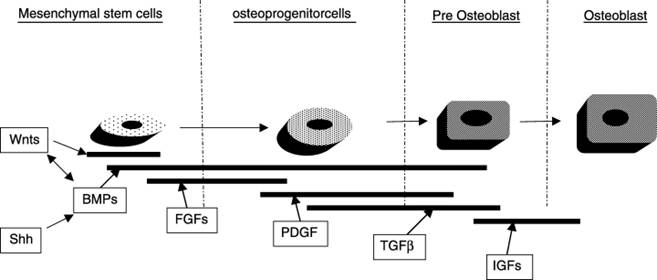
Figure217 Hughes, Francis J.; Turner, Wendy; Belibasakis, Georgios; & Martuscelli, Gianluca (2006) Effects of growth factors and cytokines on osteoblast differentiation. Periodontology 2000 41 (1), 48-72.
|
|
Fig. 2. Simplified schematic diagram showing the main stages of the osteoblast lineage where different growth factors may act. BMP, bone morphogenetic protein; IGF, insulin-like growth factor; FGF, fibroblast growth factor; PDGF, platelet-derived growth factor; Shh, Sonic hedgehog; TGF-β, transforming growth factor-β; Wnts, a group of >15 related extracellular signaling molecules.