Digital radiography has existed in the medical field for a number of years, although it is still relatively new in the dental marketplace. Like many other technologies the number of suppliers offering systems appears to be mushrooming.
Myth Number 1 - XYZ Is The Right System For Every Practice and All Digital Imaging Needs.
The digital radiography requirements of individual practices vary as much as the practices themselves. If the converse were true there would only be one successful manufacturer of dental equipment, and only one company would be offering practice management software. For this reason it is important to either select a company that can offer a full range of products or examine individual products from various suppliers.
Myth Number 2 - ABC Sensor system (Direct Digital) or DEF Phosphor Plate System (Indirect Digital) Is the Only Answer for Your Practice.
It is important to understand the benefits and drawbacks of the two types of system.
Direct digital or CCD (charge coupled device) wired sensors utilise a fibre optic sensor sandwich, which is stimulated by X-ray exposure. This area is then converted to a digital signal and transmitted (normally by cable) to the PC where via a proprietary capture card the signal is reassembled into an image.
With most systems, at least two sensors will be required to carry out day-to-day routine procedures. The major benefits, however, come with the image being immediately available on screen and elimination of the problems that can be caused through processing faults. The major drawback with this type of system is the cost involved if X-rays need to be taken in multiple surgeries on a regular basis.
At the very least the sensors must be easily transportable, as the cost of equipping each surgery with its own set of sensors can be prohibitive. Not only would sensors be required for each surgery, but also it might well prove necessary to upgrade the existing terminals and add the appropriate capture card to each surgery. The sharing of sensors between surgeries is unlikely to be a viable option in many cases.
PSP (Photostimulable Phosphor Plate Systems) photosensitive plates, not dissimilar in appearance to conventional film, are stimulated by exposure to X-rays. These are then placed in a machine and scanned with a laser. This reflected image is converted to digital information and reassembled in the PC as an image.
Unlike the CCD technology this process is not instantaneous but takes around forty five seconds for an intraoral image to display on screen. The images can then be manipulated by software as with CCD systems. The major advantages of this technology are the reduced cost for multi-surgery systems and the ease of positioning of the plates, as wired sensors can be difficult to position especially when accessing `distal' teeth.
A major drawback is the time taken to process the plates. Especially in a busy practice, where the scanner has replaced the manual processor bottlenecks can occur. It is not possible with PSP systems to commence scanning new plates until the previous scan process has been finished. Obviously with certain procedures e.g., endodontic, where the instant image is a major reason for going digital, it makes little sense to substitute a chemical processor for an electronic scanner.
Myth Number 3 - Dedicated/Special X-ray Equipment Is Required.
It has been suggested that only certain types of X-ray equipment allow you to take digital images. In fact, any machine will allow digital radiography equipment to be used.
Obviously, the newer machines with digital timers allow for greater dosage reduction, but a lot can also depend on X-ray technique. In the final analysis, if you are not getting satisfactory images with your existing equipment, merely changing to a digital system is unlikely to make a significant difference.
It is important to be aware of the new National Radiological Protection Board regulations and the various European Community regulations, which are expected to affect the legality of your system from 1999 onwards. This is particularly relevant, as it appears that this will also affect the computers you use in surgery if they are linked to diagnostic radiographic equipment. Is your proposed supplier aware of these regulations, and does the supplier have the relevant knowledge and/or contacts to advise on the suitability/legality of your existing/proposed X-ray equipment?
Myth Number 4 - Digital Radiography is More Expensive.
An interesting calculation relates to the costs of film, chemicals and mounts_savings related to film, chemicals etc. and the costs that will be levied for the disposal of chemical waste. These are only the direct costs, which are fairly straightforward to calculate. More difficult to quantify but of equal importance are the costs involved with staff time, waiting for X-rays to be developed and on occasion the necessity to repeat the procedure because the image is not of a satisfactory quality.
Myth Number 5 - Techno Blurb
ABC is the best system because it has 192 left-handed widgets. Whilst it is not uncommon to produce all sorts of meaningless data relating to a specific product, the final result must be in the quality of the images produced on screen.
Whilst line pairs are of some importance, it is equally important to understand that a number of other factors influence the quality of the final image.
The ability of the software to filter `noise' and the use of algorithms to allow easier viewing of different features can provide invaluable aids to diagnosis. It is important to ensure that the software supplied is easy to use. There is little point in saving a few minutes processing time if you are still attempting to come to grips with the software six months later and are unable to fully utilise all the features of the software.
Whilst some manufacturers claim up to 90% reduction in radiation dosage, in practice between 60 and 75% will normally be achieved per exposure, although this is in part dependent on the equipment being used and also on your expectations of how the image should look prior to any manipulation.
The facts_All digital radiography systems offer the following benefits:
Images can be stored on a hard disk or other media, thus moving the practice towards a truly `paperless'
environment.
| All systems allow for interesting manipulation of images. The ability to convert images from negative to positive
and back can sometimes assist in image enhancement. The ability to change contrast, brightness and sharpness are the
essence of digital imaging. Calibrated tissue density and length measurements are also excellent features. However,
medical and dental radiographs must be tamper proof i.e., the software should not allow you to make any permanent
changes. Of course, it will always be possible to delete files, but the unscrupulous practitioner could also destroy a
conventional X-ray.
| There is no doubt that in the foreseeable future, it will be commonplace to transmit both radiographic and intraoral
images. Do ensure that your supplier is aware of the developments in this field and has the necessary expertise to help
in this area if and when required.
| All systems allow cost savings on chemicals, films, mounts etc., see Myth Number 4. | Any system should be capable of integration into any full clinical, practice management software package.
Integration, of course, means a single database, and access to X-ray images directly from the patient's clinical record. | |
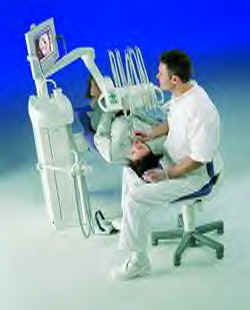
In recent months, the term `clinical workstation' has begun creeping into the vocabulary of high-tech dental equipment suppliers. So what are such companies talking about? They are referring to a surgery-based computer which runs practice management software (ideally networked throughout the practice) together with intra-oral imaging and digital radiography software and hardware. Indeed, certain companies are already marketing the clinical workstation concept. In the future we can expect to see such computer systems being fully integrated with the dental chair. Although it may be some years before this becomes the norm, it is well to be aware of the trends.

By far the most important factor in the final choice of equipment, regardless of how much of the new technology you want at this point, is to choose a supplier who knows and understands the various technologies, can offer demonstrable good support and back-up, and who will be able to assist with the integration of new technologies as they come on stream.
Source: Advanced Healthcare Imaging Limited. Photograph courtesy of Planmeca.
www.advancedhealthcareimaging.co.uk
![]() Previous Chapter
(Chapter 2)
Previous Chapter
(Chapter 2)
![]() Next Chapter
(Chapter 4)
Next Chapter
(Chapter 4)