Digital Radiography–
Not If But When
By Jack D. Preston, DDS
Abstract:
Digital radiography can enhance the dental practice by facilitating diagnosis, enabling orderly filing and
archiving, and allowing better communication with patients. Although the initial investment in equipment is substantial,
it is quickly repaid and provides both a substantive and fiscal benefit. There are challenges involved in
implementation, but they are quickly being overcome. It is only logical for dentistry to move along with the rest of
society into the digital age and take advantage of its benefits.
Change is never comfortable. Dentists spend years learning to do something well, and then a new technique comes along
and challenges the comfort of the status quo. It happens with resin composites, new bonding agents, new formulations yet
it is hard to give up something that seems to be working sufficiently. A new impression material may or may not produce
better results. While change is not comfortable, it must be recognized as inevitable. One either goes forward or falls
behind there really is no status quo. Today's dentist is faced with a myriad of changes and decisions, many of them
induced by the electronic revolution. Among the decisions to be made is whether to seriously consider digital
radiography.
Digital radiography is becoming increasingly more common, and the interested practitioner is faced with a variety of
choices and decisions. More than 10 systems are available, all offering different features. While digital radiography
has not yet become commonplace, many dentists have expressed interest in purchasing a system. The basic decisions are
whether to make the move to digital radiographs and, once the first decision is made, which digital radiographic system
to purchase.
Reasons not to move from film-based radiography to digital imaging are no longer valid for most practitioners. The
most-often-cited reasons for such reluctance have been image quality and cost. Both of these now favor digital
radiography. A third reason has been technophobia, a general resistance to entering the digital world. Perhaps this is
the issue that should be addressed first.
The Digital World
Children today are growing up in a digital world. They play with computerized toys, surf the Internet, and do their
homework on computers. To the modern generation of children, the computer, with all its functions and interconnections,
is just another accepted element of normal life. Computer software augments traditional learning methods. It broadens
and facilitates communication. The computer is becoming commonplace in the home, the school, and in business. To many
adults, however, the computer and computerized applications are still intimidating; and many are unwilling to face the
learning curve necessary to become part of modern society. Such attitudes must change if the dentist is to remain
technically competitive and be able to take advantage of devices such as digital radiography.
Consider the world in which we live. Supermarkets speed checkout using computer-based scanners and track customers'
marketing preferences in the process. Airline and hotel reservation systems are computer-based, and travelers routinely
make their own reservations from their computers. Investors rely on stock markets' use of computers to conduct business
and can track the progress of their investments on their personal computers. The Internet has proven to be a rich and
deep resource for learning, shopping, and communicating. Every substantial business is either using Internet marketing
or investigating how to do so. All levels of government, transportation, commerce, and communication rely on computers.
Even automobiles are dependent on computer processing. There would be no concern about the potential "Y2K"
problem if computers were not ubiquitous in society. Whether we choose to recognize it or not, we all live in a digital
world.
It is not a matter of if one is going to move to a digitally based office, but when. If one chooses to remain
technophobic and not make the transition to the modern world, it is a conscious choice to be left behind, to stand out
as an anachronistic relic of the past. The decision to be made, then, is how to effectively implement digital technology
into a dental practice.
Image Quality
When digital radiography was introduced into dentistry, the quality of the images was less than desirable.
Furthermore, sensors were bulkier but had active surface areas smaller than that of traditional film. Users were forced
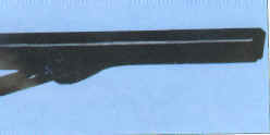
to accommodate these limitations. This has changed. Today sensors are available that are the equivalent of the commonly
used film surface area and have a comfortable thickness (Figure 1). Several digital radiography systems offer sensors in
sizes commensurate with the surface area of traditional film, including No. 0 for pediatric dentistry, No. 1 for
anterior periapical images, and No. 2, the universally used size for most imaging. The physical dimensions are easy to
compare, but comparing diagnostic quality is more difficult.
When comparing a digital image with film image, it is generally assumed that the film image has been acquired by
selecting the appropriate voltage, amperage and time, and that the properly exposed film was also properly processed
(including adequate fixation and drying) and appropriately filed. Such films are rarely found in the real world. Film is
commonly viewed without magnification and evaluated using only ambient light. If a film image was not properly exposed
or processed, and does not have optimum contrast and brightness, the dentist is forced either to make a new image or to
accept a less than desirable image. Because of time constraints, the latter choice is usually made. However, a digital
image can be manipulated to compensate for less-than-optimal exposure variables and is always viewed in a larger format
than film. Furthermore, it can be greatly magnified (the extent of the maximum magnification is dependent on the
inherent pixel size of the sensor) and can be otherwise optimized without sacrificing original image integrity. Contrast
and brightness can be varied to focus on the different features in the image.
|
|
|
Figure 1.
Modern sensors are less bulky and more comfortable than their predecessors. The sensor shown is 3.2 mm thick.
|
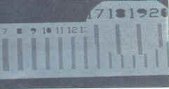
Figure 2.
A radiograph of a line pair phantom. The magnified inset shows that at 20 line pairs/mm. the separation of lines and
spaces is still clearly visible.
|
Quantifying Image Quality
The term resolution is often used to compare film and digital images. Resolution measurements attempt to quantify the
smallest observable details. It is often referred to in terms of "line pairs" the use of an imaging instrument
having successively smaller pairs of radiopaque lines. Direct digital imaging (as opposed to phosphor plate technology
that has lower resolution) offers resolution from about 11 to more than 20 line pairs (Figure 2) while film is generally
considered to resolve 15 line pairs. The unaided human eye can only see 9 line pairs, but the enhanced resolution
becomes a factor upon magnification. Sensor resolution greater than 20 line pairs has only recently been possible.
Resolution is only one aspect of the diagnostic quality of either film or digital images. The large format of the
digital image makes visualization easier and enhances communication with patients. Once one becomes accustomed to
viewing images on a large monitor rather than peering at a small film on a view box, it is difficult to return to film.
Dynamic range the blackness of blacks, whiteness of whites, and continuous tone of a complete gray scale is essential
for adequate diagnosis. Nearly all direct digital systems provide 256 grays, the greatest number the human eye is
capable of distinguishing.
Signal-to-noise ratio quantifies the strength of the signal to the background electronic noise. When magnified, film
is quite "noisy." Digital radiography systems use signal amplification and background noise subtraction to
produce the cleanest possible image.
Pixel Density
Sensors are made up of a series of electronic "wells" that trap electrons. The number of electrons in each
well translates into gray-scale images. The size of these wells varies with different sensors. A typical sensor will
have electron wells approximately 45 mm square. Unlike film, where crystals are randomly arranged and of various sizes,
the sensor has ordered rows and columns of electron wells. Each of these is an element in the image that is produced: a
picture element, or a "pixel." The smaller the pixel, the greater the number of points of information that can
be displayed on a screen. High-resolution sensors with pixels one-fourth the typical size are now available. Four pixels
of 22.5mm will fit into the space occupied by a 45 mm -square pixel. Therefore, the pixel density of the high-resolution
systems is four times greater than that of the standard systems. This will manifest as a finer grain image but will not
be greatly visible until the image is magnified. High-resolution images may be magnified more greatly without breaking
up into visible pixels (pixelating). Sensors with more than a million pixels (megapixel sensors) are available from at
least two sources, and others are sure to follow.
Regardless of what technical methods are used to make a comparison, probably the only means of really equating the
diagnostic equivalency or superiority of digital images comes from the daily use of film and sensors and appreciating
the quality of today's digital diagnostic systems.
Cost
Once diagnostic equivalency has been shown, the second factor to address is cost. Film carries both direct and
indirect costs. The cost of the film itself, the cost of processing chemicals and space in which to use them, the cost
of waste disposal, the cost of automatic film processors, and, not insignificantly, the time it takes to process and
store film all must be considered. Add to this the time required for cleanup and maintenance, and it becomes apparent
that the time consumed by film radiography is significant. Rather than construct a scenario that might or might not
apply to the reader, it is suggested that each reader calculate the number of films made each day in his or her office;
the time consumed by the assistant, hygienist, or dentist in processing, mounting, and cleaning; and especially the cost
of waiting with a patient for a film to be processed so a procedure can continue. Endodontics, implant dentistry, dowel
post placement, oral surgery, and similar services may all require sequential imaging during a procedure, and waiting
time can be substantial. Using these numbers, it is easy to demonstrate that the cost of digital X-ray system can be
recouped in less than a year with many added benefits.
With digital radiography, there are no chemicals to purchase, store, or dispose of. There is no film to carry in
inventory. There are no mounting procedures and no filing or loss of images. One of the great advantages of digital
radiography is the orderly filing of images, with the ability to retrieve by tooth, region, or date.
Radiation Reduction
Virtually everyone realizes that digital radiograph images require less radiation to make than film. The decrease in
radiation burden is usually cited as a comparison to D speed film, since the comparison is more favorable. When the
purpose of the film is to establish some gross feature, such as the position of a file in a canal, or the length of a
dowel post, a radiation reduction of up to 90 percent over D speed film is possible. For more discriminating diagnosis,
evaluating margins or small carious lesions, higher exposures are needed; but in any event, the radiation dose will be
less than either D or E speed film.
What is important is the latitude of the system. Over how wide a range can an acceptable image be made? With a wide
latitude, it is simpler to get a diagnostic quality image. It is better to err on the side of underexposure, since
overexposure may cause "burnout" loss of image information as a result of electrons spilling out of a
capture well into adjacent areas. With slight overexposure, the majority of the film will be of diagnostic quality, but
some areas will be unusable.
Remaking Images
There is another issue related to radiation reduction, and that is the re-exposure to obtain the desired image.
Sometimes an image must be remade, either because the film or sensor has been mispositioned, or the X-ray cone was
misdirected. Remakes may be necessitated with either film or sensors. However, when remaking a film image, the film has
been removed from the mouth, several minutes have passed since it was exposed, and the chances of making a proper image
the second time are not greatly better than they were with the first exposure. With a digital image, the image is
displayed almost immediately, the sensor is still in position, and the X-ray tube head is still in place. Remaking the
image becomes much more predictable, since the sensor or tube head can be moved from a known position to the desired
position.
Image Transmission
One of the great advantages of digital radiography is the ability to export images. This is an advantage not only for
sending radiographs to third-party payers but for many other purposes. Duplicate radiographs–whether single images or
a complete mouth series, have the same quality as the originals–at no added cost, and with virtually no additional
time. These can be sent to a referring dentist or forwarded to any other treating dentist.
Legal Issues
Concerns have been raised about the legal aspects of a virtual image, and the potential for image alteration
(falsification). There are now image-tagging algorithms that mark images as being original and unaltered. Such images
may be enhanced (brightness, contrast, etc.) but may not have any alteration of the image information. Such images may
be transmitted and used with assurance that, as long as the image tag guarantees the image is unaltered, the image is
secure. Of course, neither film nor virtual images can preclude the outright fraud of sending images for different
patients or other flagrant falsification.
High-Speed Communication Services
When transmitting multiple images, data transfer speed becomes an issue. Whereas "plain old telephone
service", commonly referred to as "POTS," may be adequate for small files and infrequent transmission, if
larger files are frequently sent, the office should consider other communications systems. Urban areas have many
options, including Integrated Services Data Transmission (ISDN), Symmetric Digital Signal Lines (SDSL), and cable.
Whereas POTS is able to send messages at no greater than a nominal 56 kilobits/second (actual transmission can be much
slower), SDSL and cable can send and receive 1.5 million kilobits per second. SDSL and cable service may cost as little
as $30 per month and provide Internet service 24 hours a day, seven days a week. Such service is economical, especially
when compared with some telephone company services such as a T-1 line. Asymmetric Digital Signal Lines (ADSL) and
satellite transmission offer only one-way high-speed transmission—to the office, but not upstream from the office.
Satellite may be an economical alternative in areas where the preferred services are not available, but input data will
only move at the available modem speed.
With proper software, digital images and files can be accessed from a home computer if an emergency arises. Images
may also be sent via the Internet as attachments. The destination for the image is irrelevant. It can be across the hall
or across an ocean.
Similarly, images can be printed in a letter to patients or in communication for referral. Some specialists,
especially endodontists, routinely send back images of pre- and postoperative radiographs embedded in a referral letter.
Some digital radiography software facilitates such communication by linking to form letters that can be written with
images appended directly from the original program.
Image Importing and Exporting
Modern digital radiography software allows the user to import images from attached devices such as a scanner or a
digital camera. In this way, film radiographs may be accurately scanned, enhanced when necessary, and digitally filed as
a part of the patient's virtual chart. Other documents such as prescriptions, medical reports, or laboratory work
requests can also be scanned and filed. Intraoral camera images may likewise be archived with the radiographs in a
patient chart, graphically chronicling the patient's treatment and documenting progress.
Configuration
Most dentists who do not have a computer network already installed envision beginning with digital radiography as a
cart-based or portable system. The probability of a digital radiography cart being pushed from room to room is not
great. Historically, those who bought intraoral cameras on a cart either did not use them or eventually supplied all the
operatories with cameras.
The dentist who wants to test digital radiography may well start with a cart, but it is unlikely that the cart will
be frequently moved. Another way to set up a mobile system is to use a laptop computer. Several such systems are
available, using either PCMCIA slots or universal serial bus (USB) ports. Such systems make movement from room to room
fairly easy, and the large hard drives on modern notebooks makes image storage reasonable for the short term. However,
once images are acquired and patient folders begin to accumulate, the serious user must consider networking and linking
to a practice management and virtual patient record system. It makes no sense to isolate patient X-ray images on a
computer that is not interfaced with other records. In an office where the hygienist or other associates will also be
using the system, isolation is unthinkable. In such situations, a digital network is a reasonable, viable solution. Once
computers in the different operatories are linked, patient files can be shared and are easily accessible by everyone
authorized to view them. When a practice management system and virtual patient treatment record are added, the full
facility for acquiring and archiving information is present.
The thought of integrating all records is intimidating to many dentists, and the idea of linking some other software
to their practice management system brings looks of horror and disbelief. It is true that in the past such integration
has been problematic and unreliable with some systems; but, with well-written software and a stable network, such
integration can be seamless and smooth.
When considering a network installation, one should remember that image transmission requires greater bandwidth
(ability to pass information) than does text alone. Fortunately, networking costs, as most computer-related costs, have
dropped; and a high-speed (100 Base T) network is the only reasonable consideration.
Image Size
As previously mentioned, high-resolution images have greater pixel density. This means larger file sizes, which
affect storage capabilities. Since the legality of image compression is still unclear, it is safest to opt for loss less
compression, in which no image information is discarded. Some digital systems have image storage capabilities that
guarantee an original image but at the expense of maintaining a larger file. Fortunately, hard disk sizes continue to
grow while costs drop. In planning storage capabilities, one should take into consideration the average number of
radiographs made weekly, the file size for each image, and the total file requirement to store images each year.
Archiving
Since X-ray images are a part of the patient's dental record, they must be archived. If one calculates the number of
patients treated each year, and the number of images that each patient represents, simple multiplication will indicate a
data storage problem. Images not in active use may be archived on CD-ROM, digital tape, or another server. Before long,
off-site digital storage will be available at a reasonable cost and with easy access. When high-speed data transmission
is coupled with off-site digital storage, the data storage and access problem is nicely solved.
Image Capture
Part of the intimidation factor of progressing from film to digital radiography is the change in both format and
function. The procedures for obtaining film images are so routine that there may be some reluctance to try anything that
would be more demanding. Fortunately, many current systems simplify image acquisition with little deviation from current
film-based procedures. The first noticeable difference between film and sensors is that sensors are rigid: They cannot
be bent. Operators frequently bend film to accommodate oral conditions, with resultant image distortion. Thus, the
rigidity of the sensor can be thought of as a positive factor in that it prevents distortion by bending, even though it
requires accommodation to some oral conditions.
Sensors also require a barrier cover, since they cannot be autoclaved. Some sensors are provided with plastic
barriers, others with natural rubber. The barrier should cover both the cable and the sensor–anything that contacts
the patient. Some attention should also be given to the receptacle in which the sensor will rest between uses, inasmuch
as this is also a potential source of cross-contamination. A disposable insert is desirable.
Sensor positioners should accommodate either a paralleling technique or a bisected angle approach. Positioners should
also allow for root canal instruments to remain in place during the imaging. In short, the system should allow the user
to continue the same practices currently used with film.
Image capture should involve as little interaction with the computer as possible. Some systems are able to sense the
radiation and display the image after being initially activated, minimizing any computer interfacing. The great benefit
of digital images is the ability to immediately display the image, whether it is a single image or part of a complete
mouth series. The user may choose to accept the image or correct the alignment and remake it.
Once the procedure for image acquisition is learned, users usually find it as simple as using film, without waiting
for the image, and with no need to develop, fix, or dry the image.
Image Enhancement
Digital radiography software systems offer a plethora of image manipulation algorithms, some of which are rarely
used. There are many advantages of image enhancement that should be mentioned. The most common is alteration of contrast
and brightness. Figure 3 shows an image that was underexposed but which was easily and quickly manipulated to improve
diagnostic quality.
Magnification is another frequently used feature. Instead of holding a film up to a light box and using loupes or a
magnifying glass to see more detail, digital images may be magnified simply and easily. The pixel density of the image
determines the degree of magnification possible without the image breaking up into pixels. High resolution images with
small pixels permit higher magnification. Figure 4 shows an area of an image enlarged two times, yet the visualization
is greatly enhanced.
Colorization is frequently shown by those demonstrating digital radiography, but it is usually of only casual
interest for diagnostic purposes. Colorized images are created by assigning a color to a range of grays, and the process
actually discards some information. However, colorization can be helpful in defining soft tissue and, when combined with
image inversion (reversing black and white), often shows trabecular patterns remarkably well (Figure 5).
Other combinations of features can also be helpful. Measurements are often needed when performing endodontic therapy
or placing a dowel post. Nearly all digital systems facilitate measurement. Measurements can not only be linear, but can
also navigate root curvatures. Spot-enhancing the area accentuates contrasts and aids visualization. Figure 6a shows a
file in a canal and short of the apex; Figure 6b shows the apex spot-enhanced, and the measurement made from the stop to
the tip of the file. Figure 6c depicts the spot- enhancement with the distance from the file tip to
the apical foremen. Such measurements are difficult with film. In addition to having to wait for the film to be
processed, the potential for measurement error is greater.
Image enhancement can greatly facilitate both diagnosis and treatment procedures. Digital radiography also improves
communication, since the patient can be shown the large image and can see features impossible to appreciate with film.
Text labeling, drawing on the image, and other communication devices help patients to visualize problems and understand
the necessity of therapy.
|
|
|
Figure 3a.
This image was underexposed.
|
Figure 3b.
A simple change of contrast and brightness produces an image that is of diagnostic quality.
|
|
|
|
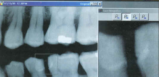
Figure 4.
The rectangle in the main image identifies the region of interest. The inset "picture-in-picture" is a 2x
magnification of the region. In reality, this image would be at least the size of this journal page, rather than the
small format shown.
|
Summary
Digital radiography facilitates and enhances diagnosis, enables orderly filing and archiving, and allows better
communication with patients. Although the initial investment may seem substantial, that sum is repaid within the first
year of use and actually provides both a substantive and fiscal benefit. It is only logical for dentistry, along with
the rest of society, to move into the digital age and take advantage of the profound benefits that the virtual world
offers. The question the practitioner should be asking is not "Why should I use digital radiography" but,
rather, "Why should I use film?"