|
4. Miscellaneous Disorders |
v Appendicitis
Although marked changes have occurred in the incidence of acute appendicitis over the century since it was first identified, the disease has changed little in its clinical features. There are no specific laboratory tests that identify it, and the multitude of other entities presenting with abdominal pain and fever ensures that as many as 20 percent of the cases operated on with the provisional diagnosis of acute appendicitis will not actually have that disease. A recent report evaluated such modalities as barium enema, ultrasound, computed tomography, and laparoscopy alone or in combination; it concluded that the use of these techniques could reduce the rate of false positive diagnosis significantly without increasing morbidity. Whether a surgeon who is certain of his or her clinical diagnosis would use them is another matter.
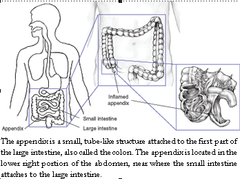
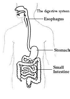
Appendicitis is an inflammation of the appendix. Once it starts, there is no effective medical therapy, so appendicitis is considered a medical emergency. When treated promptly, most patients recover without difficulty. If treatment is delayed, the appendix can burst, causing infection and even death. Appendicitis is the most common acute surgical emergency of the abdomen. Anyone can get appendicitis, but it occurs most often between the ages of 10 and 30.
|
Causes |
 |
|
The cause of appendicitis relates to blockage of the inside of the appendix, known as the lumen. The blockage leads to increased pressure, impaired blood flow, and inflammation. If the blockage is not treated, gangrene and rupture (breaking or tearing) of the appendix can result. |
|
|
Most commonly, feces blocks the inside of the appendix. Also, bacterial or viral infections in the digestive tract can lead to swelling of lymph nodes, which squeeze the appendix and cause obstruction. |
This swelling of lymph nodes is known as lymphoid hyperplasia. Traumatic injury to the abdomen may lead to appendicitis in a small number of people. Genetics may be a factor in others. For example, appendicitis that runs in families may result from a genetic variant that predisposes a person to obstruction of the appendiceal lumen.
Symptoms
Symptoms of appendicitis may include
• pain in the abdomen, first around the belly button, then moving to the lower right area
• loss of appetite
• nausea
• vomiting
• constipation or diarrhea
• inability to pass gas
• low fever that begins after other symptoms
• abdominal swelling
Not everyone with appendicitis has all the symptoms. The pain intensifies and worsens when moving, taking deep breaths, coughing, or sneezing. The area becomes very tender. People may have a sensation called “downward urge,” also known as “tenesmus,” which is the feeling that a bowel movement will relieve their discomfort. Laxatives and pain medications should not be taken in this situation. Anyone with these symptoms needs to see a qualified physician immediately.
People With Special Concerns
Patients with special conditions may not have the set of symptoms above and may simply experience a general feeling of being unwell. Patients with these conditions include
• people who use immunosuppressive therapy such as steroids
• people who have received a transplanted organ
• people infected with the HIV virus
• people with diabetes
• people who have cancer or who are receiving chemotherapy
• obese people
Pregnant women, infants and young children, and the elderly have particular issues.
Abdominal pain, nausea, and vomiting are more common during pregnancy and may or may not be the signs of appendicitis. Many women who develop appendicitis during pregnancy do not experience the classic symptoms. Pregnant women who experience pain on the right side of the abdomen need to contact a doctor. Women in their third trimester are most at risk.
Infants and young children cannot communicate their pain history to parents or doctors.
Without a clear history, doctors must rely on a physical exam and less specific symptoms, such as vomiting and fatigue. Toddlers with appendicitis sometimes have trouble eating and may seem unusually sleepy. Children may have constipation, but may also have small stools that contain mucus. Symptoms vary widely among children. If you think your child has appendicitis, contact a doctor immediately.
Older patients tend to have more medical problems than young patients. The elderly often experience less fever and less severe abdominal pain than other patients do. Many older adults do not know that they have a serious problem until the appendix is close to rupturing. A slight fever and abdominal pain on one’s right side are reasons to call a doctor right away.
All patients with special concerns and their families need to be particularly alert to a change in normal functioning and patients should see their doctors sooner, rather than later, when a change occurs.
Diagnosis
Medical History and Physical Examination
Asking questions to learn the history of symptoms and a careful physical examination are key in the diagnosis of appendicitis. The doctor will ask many questions—much like a reporter—trying to understand the nature, timing, location, pattern, and severity of pain and symptoms. Any previous medical conditions and surgeries, family history, medications, and allergies are important information to the doctor. Use of alcohol, tobacco, and any other drugs should also be mentioned. This information is considered confidential and cannot be shared without the permission of the patient.
Before beginning a physical examination, a nurse or doctor will usually measure vital signs: temperature, pulse rate, breathing rate, and blood pressure. Usually the physical examination proceeds from head to toe. Many conditions such as pneumonia or heart disease can cause abdominal pain. Generalized symptoms such as fever, rash, or swelling of the lymph nodes may point to diseases that wouldn’t require surgery.
Examination of the abdomen helps narrow the diagnosis. Location of the pain and tenderness is important. Pain is a symptom described by a patient; tenderness is the response to being touched. Two signs, called peritoneal signs, suggest that the lining of the abdomen is inflamed and surgery may be needed: rebound tenderness and guarding. Rebound tenderness is when the doctor presses on a part of the abdomen and the patient feels more tenderness when the pressure is released than when it is applied. Guarding refers to the tensing of muscles in response to touch. The doctor may also move the patient’s legs to test for pain on flexion of the hip (psoas sign), pain on internal rotation of the hip (obturator sign), or pain on the right side when pressing on the left (Rovsing’s sign). These are valuable indicators of inflammation but not all patients have them.
Laboratory Tests
Blood tests are used to check for signs of infection, such as a high white blood cell count. Blood chemistries may also show dehydration or fluid and electrolyte disorders. Urinalysis is used to rule out a urinary tractinfection. Doctors may also order a pregnancy test for women of childbearing age (those who have regular periods).
Imaging Tests
X rays, ultrasound, and computed tomography (CT) scans can produce images of the abdomen. Plain x rays can show signs of obstruction, perforation (a hole), foreign bodies, and in rare cases, an appendicolith, which is hardened stool in the appendix. Ultrasound may show appendiceal inflammation and can diagnose gall bladder disease and pregnancy. By far the most common test used, however, is the CT scan. This test provides a series of cross-sectional images of the body and can identify many abdominal conditions and facilitate diagnosis when the clinical impression is in doubt. All women of childbearing age should have a pregnancy test before undergoing any testing with x rays.
In selected cases, particularly in women when the cause of the symptoms may be either the appendix or an inflamed ovary or fallopian tube, laparoscopy may be necessary. This procedure avoids radiation, but requires general anesthesia. A laparoscope is a thin tube with a camera attached that is inserted into the body through a small cut, allowing doctors to see the internal organs. Surgery can then be performed laparoscopically if the condition present requires it.
Treatment
Surgery
Acute appendicitis is treated by surgery to remove the appendix. The operation may be performed through a standard small incision in the right lower part of the abdomen, or it may be performed using a laparoscope, which requires three to four smaller incisions. If other conditions are suspected in addition to appendicitis, they may be identified using laparoscopy. In some patients, laparoscopy is preferable to open surgery because the incision is smaller, recovery time is quicker, and less pain medication is required. The appendix is almost always removed, even if it is found to be normal. With complete removal, any later episodes of pain will not be attributed to appendicitis.
Recovery from appendectomy takes a few weeks. Doctors usually prescribe pain medication and ask patients to limit physical activity. Recovery from laparoscopic appendectomy is generally faster, but limiting strenuous activity may still be necessary for 4 to 6 weeks after surgery. Most people treated for appendicitis recover excellently and rarely need to make any changes in their diet, exercise, or lifestyle.
Antibiotics and Other Treatments
If the diagnosis is uncertain, people may be watched and sometimes treated with antibiotics. This approach is taken when the doctor suspects that the patient’s symptoms may have a nonsurgical or medically treatable cause. If the cause of the pain is infectious, symptoms resolve with intravenous antibiotics and intravenous fluids. In general, however, appendicitis cannot be treated with antibiotics alone and will require surgery.
Occasionally the body is able to control an appendiceal perforation by forming an abscess. An abscess occurs when an infection is walled off in one part of the body. The doctor may choose to drain the abscess and leave the drain in the abscess cavity for several weeks. An appendectomy may be scheduled after the abscess is drained.
Complications
The most serious complication of appendicitis is rupture. The appendix bursts or tears if appendicitis is not diagnosed quickly and goes untreated. Infants, young children, and older adults are at highest risk. A ruptured appendix can lead to peritonitis and abscess. Peritonitis is a dangerous infection that happens when bacteria and other contents of the torn appendix leak into the abdomen. In people with appendicitis, an abscess usually takes the form of a swollen mass filled with fluid and bacteria. In a few patients, complications of appendicitis can lead to organ failure and death.
Prevention
There is not enough known about its etiology to assume that dietary or other lifestyle changes will prevent appendicitis. An increase in fiber consumption has been recommended, but there are few data to recommend this approach, although it may be of benefit in the prevention of other diseases. For many years, an incidental appendectomy has been performed at the time of laparotomy for other disorders, particularly gynecologic conditions, to prevent subsequent appendicitis. A study of the epidemiology of appendectomy found that 36 incidental procedures need to be performed to “prevent” one case of appendicitis .
Points to Remember
| • | The appendix is a small, tubelike structure attached to the first part of the colon. Appendicitis is an inflammation of the appendix. |
| • | Appendicitis is considered a medical emergency. |
| • | Symptoms of appendicitis include pain in the abdomen, loss of appetite, nausea, vomiting, constipation or diarrhea, inability to pass gas, low-grade fever, and abdominal swelling. Not everyone with appendicitis has all the symptoms. |
| • | Physical examination, laboratory tests, and imaging tests are used to diagnose appendicitis. |
| • | Acute appendicitis is treated by surgery to remove the appendix. |
| • | The most serious complication of appendicitis is rupture, which can lead to peritonitis and abscess. |
v Hemorrhoids
Clinical Epidemiology of Hemorrhoids
The occurrence of hemorrhoids can be traced to antiquity. Hemorrhoids have been referred to in literature as far back as the pre-Christian era, and proctology thrived in ancient Egypt. Other ancient cultures have likewise suffered from this disorder; for instance, the Japanese, who established a temple in the City of Kanazawa dedicated to the cure of hemorrhoids. Despite their lengthy history, only recently has there been a thorough anatomic and clinical description.
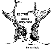
Hemorrhoids begin as localized cushions of specialized submucosal vascular tissue located adjacent to the site of the junction (the dentate line) of squamous and rectal columnar epithelium in the anal canal. Present at birth, these cushions are normal anatomical features of the anal canal. Hence, the existence of hemorrhoidal cushions alone does not constitute disease. Hemorrhoidal disease, that is, hemorrhoids, requires the presence of pathologic changes leading to bleeding, prolapse, or thrombosis. Hemorrhoids are internal or external, depending on their location in the anal canal. External hemorrhoids are located below the dentate line and are covered by squamous epithelium. In contrast, internal hemorrhoids are located above the dentate line and are covered by columnar epithelium. Internal hemorrhoids are further classified based on the extent of their prolapse. First degree hemorrhoids are enlarged but do not prolapse. Second degree hemorrhoids prolapse out of the anus with defecation but reduce spontaneously. Third degree hemorrhoids prolapse with defecation but must be reduced manually. Fourth degree hemorrhoids cannot be reduced.
What are hemorrhoids?
The term hemorrhoids refers to a condition in which the veins around the anus or lower rectum are swollen and inflamed.
Hemorrhoids may result from straining to move stool. Other contributing factors include pregnancy, aging, chronic constipation or diarrhea, and anal intercourse. Hemorrhoids are either inside the anus (internal) or under the skin around the anus (external). (See figure.)
What are the symptoms of hemorrhoids?
Many anorectal problems, including fissures, fistulae, abscesses, or irritation and itching (pruritus ani), have similar symptoms and are incorrectly referred to as hemorrhoids.
Hemorrhoids usually are not dangerous or life threatening. In most cases, hemorrhoidal symptoms will go away within a few days.
Although many people have hemorrhoids, not all experience symptoms. The most common symptom of internal hemorrhoids is bright red blood covering the stool, on toilet paper, or in the toilet bowl. However, an internal hemorrhoid may protrude through the anus outside the body, becoming irritated and painful. This is known as a protruding hemorrhoid.
Symptoms of external hemorrhoids may include painful swelling or a hard lump around the anus that results when a blood clot forms. This condition is known as a thrombosed external hemorrhoid.
In addition, excessive straining, rubbing, or cleaning around the anus may cause irritation with bleeding and/or itching, which may produce a vicious cycle of symptoms. Draining mucus may also cause itching.
How common are hemorrhoids?
Hemorrhoids are very common in both men and women. About half of the population have hemorrhoids by age 50. Hemorrhoids are also common among pregnant women. The pressure of the fetus in the abdomen, as well as hormonal changes, cause the hemorrhoidal vessels to enlarge. These vessels are also placed under severe pressure during childbirth. For most women, however, hemorrhoids caused by pregnancy are a temporary problem.
|
How are hemorrhoids diagnosed? |
|
|
|
 |
It is important that rectal bleeding not simply be attributed to hemorrhoids until other, more serious conditions are excluded. Consequently, rectal bleeding in elderly patients or in patients with atypical histories requires a complete diagnostic evaluation, even if obvious prolapsing hemorrhoids are present.
A thorough evaluation and proper diagnosis by the doctor is important any time bleeding from the rectum or blood in the stool occurs. Bleeding may also be a symptom of other digestive diseases, including colorectal cancer.
The doctor will examine the anus and rectum to look for swollen blood vessels that indicate hemorrhoids and will also perform a digital rectal exam with a gloved, lubricated finger to feel for abnormalities.
Closer evaluation of the rectum for hemorrhoids requires an exam with an anoscope, a hollow, lighted tube useful for viewing internal hemorrhoids, or a proctoscope, useful for more completely examining the entire rectum.
To rule out other causes of gastrointestinal bleeding, the doctor may examine the rectum and lower colon (sigmoid) with sigmoidoscopy or the entire colon with colonoscopy. Sigmoidoscopy and colonoscopy are diagnostic procedures that also involve the use of lighted, flexible tubes inserted through the rectum.
What is the treatment?
Medical treatment of hemorrhoids is aimed initially at relieving symptoms. Measures to reduce symptoms include
• tub baths several times a day in plain, warm water for about 10 minutes
• application of a hemorroidal cream or suppository to the affected area for a limited time
Preventing the recurrence of hemorrhoids will require relieving the pressure and straining of constipation. Doctors will often recommend increasing fiber and fluids in the diet. Eating the right amount of fiber and drinking six to eight glasses of fluid (not alcohol) result in softer, bulkier stools. A softer stool makes emptying the bowels easier and lessens the pressure on hemorrhoids caused by straining. Eliminating straining also helps prevent the hemorrhoids from protruding.
Good sources of fiber are fruits, vegetables, and whole grains. In addition, doctors may suggest a bulk stool softener or a fiber supplement such as psyllium (Metamucil) or methylcellulose (Citrucel).
In some cases, hemorrhoids must be treated endoscopically or surgically. These methods are used to shrink and destroy the hemorrhoidal tissue. The doctor will perform the procedure during an office or hospital visit.
A number of methods may be used to remove or reduce the size of internal hemorrhoids. These techniques include
| • | Rubber band ligation. A rubber band is placed around the base of the hemorrhoid inside the rectum. The band cuts off circulation, and the hemorrhoid withers away within a few days. |
| • | Sclerotherapy. A chemical solution is injected around the blood vessel to shrink the hemorrhoid. |
| • | Infrared coagulation. A special device is used to burn hemorrhoidal tissue. |
| • | Hemorrhoidectomy. Occasionally, extensive or severe internal or external hemorrhoids may require removal by surgery known as hemorrhoidectomy. |
How are hemorrhoids prevented?
The best way to prevent hemorrhoids is to keep stools soft so they pass easily, thus decreasing pressure and straining, and to empty bowels as soon as possible after the urge occurs. Exercise, including walking, and increased fiber in the diet help reduce constipation and straining by producing stools that are softer and easier to pass.
v Gallstones
Extrapolation from small studies suggests that about 20 million U.S. residents have gallstones or have had surgery for gallstones. Most persons with gallstones are asymptomatic and therefore unaware of the condition. Risk of gallstones is highest among American Indians, lower among Europeans, and lowest among black Americans and Eastern Asians. The risk of gallstones is higher among women, particularly young women, and the risk for surgery and hospitalization increases with age. Obesity is an established risk factor, particularly in women, but rapid weight loss also increases the risk. Supplemental estrogen may increase the risk modestly. The risk of gallstones dramatically increases during pregnancy and subsides shortly after delivery. High serum triglyceride concentration and low high-density lipoprotein (HDL) cholesterol concentration increase the risk of gallstones, but total serum cholesterol concentration does not. Surprisingly, little effect of dietary constituents has been found on gallstone disease, but high fiber and moderate alcohol intake may be protective; a prolonged fasting period may increase the risk. Cigarette smoking has consistently been associated with gallstones, but without obvious explanation.
Diet and Alcohol
There seems to be a common assumption that diet and nutrient composition play strong roles in the development of gallstones. With extreme dietary manipulation this may be true in some animals, and in humans dietary modification may modify biliary cholesterol saturation. Nevertheless, it has been difficult to establish an association between dietary factors and the presence of gallstones in humans. Total dietary fat and dietary cholesterol do not appear to have a strong association with gallstone disease, while associations with total energy have been remarkably inconsistent; energy intake and gallstone disease have been found to be positively related, negatively related, unrelated, and either positively or negatively, related in persons younger but not older than age 50 years. Possibly the most consistent association has been a negative association with fiber intake, but even this has not always been found. Energy deficiency resulting in weight loss clearly increases the risk of gallstones, and there is evidence that long overnight fasting also increases gallstone risk.
Moderate alcohol intake has been associated with lower risk of gallstones in several studies. Increased HDL cholesterol is associated with moderate alcohol intake, but it would be speculative to state that the lower risk of gallstones with alcohol consumption was being mediated by a specific effect on lipid metabolism. On the other hand, cirrhosis of the liver, whether due to alcohol abuse or other causes, is associated with an increased prevalence of gallstones.
|
|
What are gallstones?
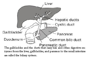
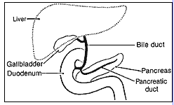
Gallstones form when liquid stored in the gallbladder hardens into pieces of stone-like material. The liquid, called bile, is used to help the body digest fats. Bile is made in the liver, then stored in the gallbladder until the body needs to digest fat. At that time, the gallbladder contracts and pushes the bile into a tube–called the common bile duct–that carries it to the small intestine, where it helps with digestion.
Bile contains water, cholesterol, fats, bile salts, proteins, and bilirubin. Bile salts break up fat, and bilirubin gives bile and stool a yellowish color. If the liquid bile contains too much cholesterol, bile salts, or bilirubin, under certain conditions it can harden into stones.
The two types of gallstones are cholesterol stones and pigment stones. Cholesterol stones are usually yellow-green and are made primarily of hardened cholesterol. They account for about 80 percent of gallstones. Pigment stones are small, dark stones made of bilirubin. Gallstones can be as small as a grain of sand or as large as a golf ball. The gallbladder can develop just one large stone, hundreds of tiny stones, or almost any combination.
The gallbladder and the ducts that carry bile and other digestive enzymes from the liver, gallbladder, and pancreas to the small intestine are called the biliary system.
Gallstones can block the normal flow of bile if they lodge in any of the ducts that carry bile from the liver to the small intestine. That includes the hepatic ducts, which carry bile out of the liver; the cystic duct, which takes bile to and from the gallbladder; and the common bile duct, which takes bile from the cystic and hepatic ducts to the small intestine. Bile trapped in these ducts can cause inflammation in the gallbladder, the ducts, or, rarely, the liver. Other ducts open into the common bile duct, including the pancreatic duct, which carries digestive enzymes out of the pancreas. If a gallstone blocks the opening to that duct, digestive enzymes can become trapped in the pancreas and cause an extremely painful inflammation called gallstone pancreatitis.
If any of these ducts remain blocked for a significant period of time, severe–possibly fatal–damage or infections can occur, affecting the gallbladder, liver, or pancreas. Warning signs of a serious problem are fever, jaundice, and persistent pain.
What causes gallstones?
Cholesterol Stones
Scientists believe cholesterol stones form when bile contains too much cholesterol, too much bilirubin, or not enough bile salts, or when the gallbladder does not empty as it should for some other reason.
Pigment Stones
The cause of pigment stones is uncertain. They tend to develop in people who have cirrhosis, biliary tract infections, and hereditary blood disorders such as sickle cell anemia in which too much bilirubin is formed.
Other Factors
It is believed that the mere presence of gallstones may cause more gallstones to develop. However, other factors that contribute to gallstones have been identified, especially for cholesterol stones.
| • | Obesity. Obesity is a major risk factor for gallstones, especially in women. A large clinical study showed that being even moderately overweight increases one’s risk for developing gallstones. The most likely reason is that obesity tends to reduce the amount of bile salts in bile, resulting in more cholesterol. Obesity also decreases gallbladder emptying. |
| • | Estrogen. Excess estrogen from pregnancy, hormone replacement therapy, or birth control pills appears to increase cholesterol levels in bile and decrease gallbladder movement, both of which can lead to gallstones. |
| • | Ethnicity. Native Americans have a genetic predisposition to secrete high levels of cholesterol in bile. In fact, they have the highest rate of gallstones in the United States. A majority of Native American men have gallstones by age 60. Among the Pima Indians of Arizona, 70 percent of women have gallstones by age 30. Mexican American men and women of all ages also have high rates of gallstones. |
| • | Gender. Women between 20 and 60 years of age are twice as likely to develop gallstones as men. |
| • | Age. People over age 60 are more likely to develop gallstones than younger people. |
| • | Cholesterol-lowering drugs. Drugs that lower cholesterol levels in blood actually increase the amount of cholesterol secreted in bile. This in turn can increase the risk of gallstones. |
| • | Diabetes. People with diabetes generally have high levels of fatty acids called triglycerides. These fatty acids increase the risk of gallstones. |
| • | Rapid weight loss. As the body metabolizes fat during rapid weight loss, it causes the liver to secrete extra cholesterol into bile, which can cause gallstones. |
| • | Fasting. Fasting decreases gallbladder movement, causing the bile to become overconcentrated with cholesterol, which can lead to gallstones. |
Who is at risk for gallstones?
• women
• people over age 60
• Native Americans
• Mexican Americans
• overweight men and women
• people who fast or lose a lot of weight quickly
• pregnant women, women on hormone therapy, and women who use birth control pills
What are the symptoms?
Symptoms of gallstones are often called a gallstone “attack” because they occur suddenly. A typical attack can cause
• steady pain in the upper abdomen that increases rapidly and lasts from 30 minutes to
several hours
• pain in the back between the shoulder blades
• pain under the right shoulder
• nausea or vomiting
Gallstone attacks often follow fatty meals, and they may occur during the night. Other gallstone symptoms include:
• abdominal bloating
• recurring intolerance of fatty foods
• colic
• belching
• gas
• indigestion
People who also have the above and any of following symptoms should see a doctor right away:
• sweating
• chills
• low-grade fever
• yellowish color of the skin or whites of the eyes
• clay-colored stools
Many people with gallstones have no symptoms. These patients are said to be asymptomatic, and these stones are called “silent stones.” They do not interfere in gallbladder, liver, or pancreas function and do not need treatment.
How are gallstones diagnosed?
Many gallstones, especially silent stones, are discovered by accident during tests for other problems. But when gallstones are suspected to be the cause of symptoms, the doctor is likely to do an ultrasound exam. Ultrasound uses sound waves to create images of organs. Sound waves are sent toward the gallbladder through a handheld device that a technician glides over the abdomen. The sound waves bounce off the gallbladder, liver, and other organs such as a pregnant uterus, and their echoes make electrical impulses that create a picture of the organ on a video monitor. If stones are present, the sound waves will bounce off them, too, showing their location. Ultrasound is the most sensitive and specific test for gallstones.
Other tests used in diagnosis include:
| • | Computed tomography (CT) scan may show the gallstones or complications. |
| • | MR cholangiogram may diagnose blocked bile ducts. |
| • | Cholescintigraphy (HIDA scan) is used to diagnose abnormal contraction of the gallbladder or obstruction. The patient is injected with a radioactive material that is taken up in the gallbladder, which is then stimulated to contract. |
| • | Endoscopic retrograde cholangiopancreatography (ERCP). The patient swallows an endoscope—a long, flexible, lighted tube connected to a computer and TV monitor. The doctor guides the endoscope through the stomach and into the small intestine. The doctor then injects a special dye that temporarily stains the ducts in the biliary system. ERCP is used to locate and remove stones in the ducts. |
| • | Blood tests. Blood tests may be used to look for signs of infection, obstruction, pancreatitis, or jaundice. |
Gallstone symptoms are similar to those of heart attack, appendicitis, ulcers, irritable bowel syndrome, hiatal hernia, pancreatitis, and hepatitis. So accurate diagnosis is important.
What is the treatment?
Surgery
Surgery to remove the gallbladder is the most common way to treat symptomatic gallstones. (Asymptomatic gallstones usually do not need treatment.) Each year more than 500,000 Americans have gallbladder surgery. The surgery is called cholecystectomy.
The most common operation is called laparoscopic cholecystectomy. For this operation, the surgeon makes several tiny incisions in the abdomen and inserts surgical instruments and a miniature video camera into the abdomen. The camera sends a magnified image from inside the body to a video monitor, giving the surgeon a close-up view of the organs and tissues. While watching the monitor, the surgeon uses the instruments to carefully separate the gallbladder from the liver, ducts, and other structures. Then the cystic duct is cut and the gallbladder removed through one of the small incisions.
Because the abdominal muscles are not cut during laparoscopic surgery, patients have less pain and fewer complications than they would have had after surgery using a large incision across the abdomen.
Recovery usually involves only one night in the hospital, followed by several days of restricted activity at home.
If the surgeon discovers any obstacles to the laparoscopic procedure, such as infection or scarring from other operations, the operating team may have to switch to open surgery. In some cases the obstacles are known before surgery, and an open surgery is planned. It is called “open” surgery because the surgeon has to make a 5- to 8-inch incision in the abdomen to remove the gallbladder. This is a major surgery and may require about a 2- to 7-day stay in the hospital and several more weeks at home to recover. Open surgery is required in about 5 percent of gallbladder operations.
The most common complication in gallbladder surgery is injury to the bile ducts. An injured common bile duct can leak bile and cause a painful and potentially dangerous infection. Mild injuries can sometimes be treated nonsurgically. Major injury, however, is more serious and requires additional surgery.
If gallstones are in the bile ducts, the physician (usually a gastroenterologist) may use endoscopic retrograde cholangiopancreatography (ERCP) to locate and remove them before or during the gallbladder surgery. In ERCP, the patient swallows an endoscope—a long, flexible, lighted tube connected to a computer and TV monitor. The doctor guides the endoscope through the stomach and into the small intestine. The doctor then injects a special dye that temporarily stains the ducts in the biliary system. Then the affected bile duct is located and an instrument on the endoscope is used to cut the duct. The stone is captured in a tiny basket and removed with the endoscope.
Occasionally, a person who has had a cholecystectomy is diagnosed with a gallstone in the bile ducts weeks, months, or even years after the surgery. The two-step ERCP procedure is usually successful in removing the stone.
Nonsurgical Treatment
Nonsurgical approaches are used only in special situations—such as when a patient has a serious medical condition preventing surgery—and only for cholesterol stones. Stones usually recur after nonsurgical treatment.
| • | Oral dissolution therapy. Drugs made from bile acid are used to dissolve the stones. The drugs, ursodiol (Actigall) and chenodiol (Chenix), work best for small cholesterol stones. Months or years of treatment may be necessary before all the stones dissolve. Both drugs cause mild diarrhea, and chenodiol may temporarily raise levels of blood cholesterol and the liver enzyme transaminase. |
| • | Contact dissolution therapy. This experimental procedure involves injecting a drug directly into the gallbladder to dissolve stones. The drug–methyl tertbutyl ether–can dissolve some stones in 1 to 3 days, but it must be used very carefully because it is a flammable anesthetic that can be toxic. The procedure is being tested in patients with symptomatic, noncalcified cholesterol stones. |
| • | Extracorporeal shockwave lithotripsy (ESWL). This treatment uses shock waves to break up stones into tiny pieces that can pass through the bile ducts without causing blockages. Attacks of biliary colic (intense pain) are common after treatment, and ESWL’s success rate is not known. This approach is usually combined with therapeutic ERCP. |
Don’t people need their gallbladders?
Fortunately, the gallbladder is an organ that people can live without. Losing it won’t even require a change in diet. Once the gallbladder is removed, bile flows out of the liver through the hepatic ducts into the common bile duct and goes directly into the small intestine, instead of being stored in the gallbladder. However, because the bile isn’t stored in the gallbladder, it flows into the small intestine more frequently, causing diarrhea in about 1 percent of people.
Points to Remember
• Gallstones form when substances in the bile harden.
• Gallstones are more common among women, Native Americans, Mexican Americans, and people who
are overweight.
• Gallstone attacks often occur after eating a meal.
• Symptoms can mimic those of other problems, including heart attack, so accurate diagnosis is important.
• Gallstones can cause serious problems if they become trapped in the bile ducts.
• Laparoscopic surgery to remove the gallbladder is the most common treatment.
v Diverticulosis and Diverticulitis
|
Summary |
 |
and dietary fiber. In rapidly westernizing countries, however, there has been a rather rapid increase in the disorder. Current opinion is that the disorder occurs as a slow adaptation to diet, although there are no long-term feeding studies in humans to support this assumption. No studies have been strictly population-based, and all reports depend on contact of patients with the health care system to have definitive diagnostic procedures carried out. Almost all data available consist of cases found on routine studies of persons sick enough to complain of symptoms suggestive either of diverticulitis or of other bowel disorders that require barium or colonoscopic studies for their resolution. Most cases of diverticulosis are found incidentally because there is no evidence that diverticula produce symptoms in 85 percent of the diverticula-bearing population. Diverticulitis, however, results often in disability, occasionally in hospitalization, more rarely in surgical treatment, and in recurrent problems over the years.
Every year nearly 2 million persons report having had diverticular disease with the prevalence in the elderly nearly 40 times that of persons younger than age 45 years. There are nearly 2 million visits to office- based physicians, 440,000 hospitalizations, and about 3,000 deaths attributed to diverticular disease each year, nearly one-third among persons 85 years of age and older. Improvements in coding and the development and use of criteria for diagnosis would aid the investigation of diverticular disease.
Definitions
Diverticula are outpouchings in hollow tubes, wherein the inner layers of the tubes protrude through defects in the outer layers and may project for some distance into surrounding tissues or tissue spaces. In the gastrointestinal tract, diverticula occur under two conditions: first, when muscle fibers of one system or anatomic type mesh with muscle fibers of a differing type, producing an imperfect tissue with defects allowing for protrusion; second, when the principal musculature of the tube wall, for one reason or another, pulls away from the vascular tunnels through that wall and creates small spaces through which the mucosa can herniate and eventually protrude by lifting up the serosa of the digestive tube to form diverticula, usually small at first. Examples of the first type are the Zenker’s diverticulum in the hypopharynx, gastric diverticulum where the esophagus joins the stomach, and duodenal diverticulum at the point where the biliary and pancreatic ducts enter the duodenum. The second type, which is much more common, occasionally is seen in the small intestine, but characteristically it is found in the colon, where it most often is multiple. The term “diverticular disease” is a recent one designed to describe the clinical disorders that may be associated with the anatomic presence of diverticula. This term lacks precision, but it has been adopted because our ability to discriminate the various underlying causes for symptoms in patients whose colons contain diverticula is not very good. Symptoms ascribed to diverticular disease are similar to those associated with the irritable bowel syndrome, for which the presence or absence of diverticula seems to be unimportant.
Current Means of Diagnosis
Diverticular disease represents a relatively recent area of medical science, probably because the populations at risk in the past were much younger than the current western ones and because approaches to intestinal pathology infrequently were surgical and only in the 20th century radiological. Because many diverticula are small and the colon is covered with serosal fat, routine autopsies in the 19th century probably overlooked them unless there was a peridiverticular septic process or a fistulization between the inflamed adjacent viscera. The advent of barium contrast studies in the first decades of this century made clear the existence of the disorder and began to establish criteria for its diagnosis.
With the advent of endoscopic approaches to colonic diseases, it became clear that diverticula often were visualized during colonoscopy. Such visualizations are regarded as confirming the diagnosis, although they are not as reliable for assessing the overall extent of diverticulisis as is the barium enema. Currently, the diagnosis of diverticulosis must be validated by barium contrast studies, endoscopy, or surgery. The diagnosis of diverticulitis is made on the clinical grounds of fever, leukocytosis, localized lower abdominal pain, and change in bowel habit in a patient known to have diverticulosis. Confirmatory findings are obtained by computerized tomography, which may demonstrate localized abscess formation; by colonoscopy, which may provide evidence of localized mucosal inflammation or bleeding; or by combinations of barium studies and colonoscopy, which suggest partial colonic obstruction. Rectal bleeding alone is not a feature of diverticular disease, although in patients presenting with rectal bleeding and in whom no firm source of bleeding is established, the existence of diverticulosis often is regarded as the source. This is a serious error.
Natural History
The course of diverticular disease is asymptomatic in about 85 percent of cases. The disease may begin in the fifth decade and may be fully expressed a decade or two later, but characteristically its clinical emergence depends on a bout of peridiverticulitis, a localized peritonitis of small extent and with few findings on physical examination. The patient may complain of a change in bowel habit, usually toward constipation, and of lower abdominal pain accompanied by some low-grade fever. These findings usually lead to an examination of the colon by barium enema or colonoscopy and the diagnosis of diverticular disease. Treatment of this episode results in an illness of about 1 week. Studies following patients first diagnosed and treated for diverticular disease have estimated that about 25 percent of those diagnosed will have further bouts of peridiverticulitis over a lifetime. A much smaller number of patients will have complications of the process, leading to abscess or fistula formation and requiring major colonic surgery for treatment. The current methods of managing the acute bout have seemed to decrease markedly the massive surgical complications, the pylephlebitis, and the complications of general peritonitis that so dominated the older literature on this subject. Diverticular disease does not have known effects on other colonic diseases and is not in itself a precursor of colonic polyps or cancer, although the latter occur in the same age group and may be related dietetically.
Current ideas on the dietary management of diverticular disease have been moderately successful in reducing the frequency of recurrences, and the available antibiotics have been particularly useful in reducing the morbidity attributable to peritonitis.
Prevention
The prevention of diverticular disease may prove to be no more than the adoption of a diet containing more vegetables, fruits, legumes, and grains, and less animal fats and protein–in other words, the “prudent diet” currently promoted for the prophylaxis of heart disease and cancer. It should be pointed out that there is no direct relationship of diverticular disease to heart disease or colonic cancer. In Oriental populations, colonic polyps seem unrelated to colonic diverticulosis.
Diverticulum, diverticulosis, diverticulitis
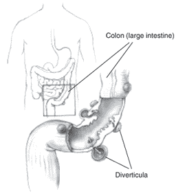
Many people have small pouches in their colons that bulge outward through weak spots, like an inner tube that pokes through weak places in a tire. Each pouch is called a diverticulum. Pouches (plural) are called diverticula. The condition of having diverticula is called diverticulosis. About 10 percent of Americans over the age of 40 have diverticulosis. The condition becomes more common as people age. About half of all people over the age of 60 have diverticulosis.
When the pouches become infected or inflamed, the condition is called diverticulitis. This happens in 10 to 25 percent of people with diverticulosis. Diverticulosis and diverticulitis are also called diverticular disease.
What causes diverticular disease?
Although not proven, the dominant theory is that a low-fiber diet is the main cause of diverticular disease. The disease was first noticed in the United States in the early 1900s. At about the same time, processed foods were introduced into the American diet. Many processed foods contain refined, low-fiber flour. Unlike whole-wheat flour, refined flour has no wheat bran.
Diverticular disease is common in developed or industrialized countries–particularly the United States, England, and Australia—where low-fiber diets are common. The disease is rare in countries of Asia and Africa, where people eat high-fiber vegetable diets.
Fiber is the part of fruits, vegetables, and grains that the body cannot digest. Some fiber dissolves easily in water (soluble fiber). It takes on a soft, jellylike texture in the intestines. Some fiber passes almost unchanged through the intestines (insoluble fiber). Both kinds of fiber help make stools soft and easy to pass. Fiber also prevents constipation.
Constipation makes the muscles strain to move stool that is too hard. It is the main cause of increased pressure in the colon. This excess pressure might cause the weak spots in the colon to bulge out and become diverticula.
Diverticulitis occurs when diverticula become infected or inflamed. Doctors are not certain what causes the infection. It may begin when stool or bacteria are caught in the diverticula. An attack of diverticulitis can develop suddenly and without warning.
What are the symptoms?
Diverticulosis
Most people with diverticulosis do not have any discomfort or symptoms. However, symptoms may include mild cramps, bloating, and constipation. Other diseases such as irritable bowel syndrome (IBS) and stomach ulcers cause similar problems, so these symptoms do not always mean a person has diverticulosis. You should visit your doctor if you have these troubling symptoms.
Diverticulitis
The most common symptom of diverticulitis is abdominal pain. The most common sign is tenderness around the left side of the lower abdomen. If infection is the cause, fever, nausea, vomiting, chills, cramping, and constipation may occur as well. The severity of symptoms depends on the extent of the infection and complications.
What are the complications?
Diverticulitis can lead to bleeding, infections, perforations or tears, or blockages. These complications always require treatment to prevent them from progressing and causing serious illness.
Bleeding
Bleeding from diverticula is a rare complication. When diverticula bleed, blood may appear in the toilet or in your stool. Bleeding can be severe, but it may stop by itself and not require treatment. Doctors believe bleeding diverticula are caused by a small blood vessel in a diverticulum that weakens and finally bursts. If you have bleeding from the rectum, you should see your doctor. If the bleeding does not stop, surgery may be necessary.
Abscess, Perforation, and Peritonitis
The infection causing diverticulitis often clears up after a few days of treatment with antibiotics. If the condition gets worse, an abscess may form in the colon.
An abscess is an infected area with pus that may cause swelling and destroy tissue. Sometimes the infected diverticula may develop small holes, called perforations. These perforations allow pus to leak out of the colon into the abdominal area. If the abscess is small and remains in the colon, it may clear up after treatment with antibiotics. If the abscess does not clear up with antibiotics, the doctor may need to drain it.
To drain the abscess, the doctor uses a needle and a small tube called a catheter. The doctor inserts the needle through the skin and drains the fluid through the catheter. This procedure is called percutaneous catheter drainage. Sometimes surgery is needed to clean the abscess and, if necessary, remove part of the colon.
A large abscess can become a serious problem if the infection leaks out and contaminates areas outside the colon. Infection that spreads into the abdominal cavity is called peritonitis. Peritonitis requires immediate surgery to clean the abdominal cavity and remove the damaged part of the colon. Without surgery, peritonitis can be fatal.
Fistula
A fistula is an abnormal connection of tissue between two organs or between an organ and the skin. When damaged tissues come into contact with each other during infection, they sometimes stick together. If they heal that way, a fistula forms. When diverticulitis-related infection spreads outside the colon, the colon’s tissue may stick to nearby tissues. The organs usually involved are the bladder, small intestine, and skin.
The most common type of fistula occurs between the bladder and the colon. It affects men more than women. This type of fistula can result in a severe, long-lasting infection of the urinary tract. The problem can be corrected with surgery to remove the fistula and the affected part of the colon.
Intestinal Obstruction
The scarring caused by infection may cause partial or total blockage of the large intestine. When this happens, the colon is unable to move bowel contents normally. When the obstruction totally blocks the intestine, emergency surgery is necessary. Partial blockage is not an emergency, so the surgery to correct it can be planned.
How does the doctor diagnose diverticular disease?
To diagnose diverticular disease, the doctor asks about medical history, does a physical exam, and may perform one or more diagnostic tests. Because most people do not have symptoms, diverticulosis is often found through tests ordered for another ailment.
When taking a medical history, the doctor may ask about bowel habits, symptoms, pain, diet, and medications. The physical exam usually involves a digital rectal exam. To perform this test, the doctor inserts a gloved, lubricated finger into the rectum to detect tenderness, blockage, or blood. The doctor may check stool for signs of bleeding and test blood for signs of infection. The doctor may also order x rays or other tests.
What is the treatment for diverticular disease?
A high-fiber diet and, occasionally, mild pain medications will help relieve symptoms in most cases. Sometimes an attack of diverticulitis is serious enough to require a hospital stay and possibly surgery.
Diverticulosis
Increasing the amount of fiber in the diet may reduce symptoms of diverticulosis and prevent complications such as diverticulitis. Fiber keeps stool soft and lowers pressure inside the colon so that bowel contents can move through easily. The American Dietetic Association recommends 20 to 35 grams of fiber each day. The table below shows the amount of fiber in some foods that you can easily add to your diet.
| Amount of Fiber in Some Foods | |
|
Fruits Apple, raw, with skin Peach, raw Pear, raw Tangerine, raw Vegetables Asparagus, fresh, cooked Broccoli, fresh, cooked Brussels sprouts, fresh, cooked Cabbage, fresh, cooked Carrot, fresh, cooked Cauliflower, fresh, cooked Romaine lettuce Spinach, fresh, cooked Summer squash, cooked Tomato, raw Winter squash, cooked Starchy Vegetables Baked beans, canned, plain Kidney beans, fresh, cooked Lima beans, fresh, cooked Potato, fresh, cooked Grains Bread, whole-wheat Brown rice, cooked Cereal, bran flake Oatmeal, plain, cooked White rice, cooked |
1 medium = 3.3 grams 1 medium = 1.5 grams 1 medium = 5.1 grams 1 medium = 1.9 grams 4 spears = 1.2 grams ˝ cup = 2.6 grams ˝ cup = 2 grams ˝ cup = 1.5 grams ˝ cup = 2.3 grams ˝ cup = 1.7 grams 1 cup = 1.2 grams ˝ cup = 2.2 grams 1 cup = 2.5 grams 1 = 1 grams 1 cup = 5.7 grams ˝ cup = 6.3 grams ˝ cup = 5.7 grams ˝ cup = 6.6 grams 1 = 2.3 grams 1 slice = 1.9 grams 1 cup = 3.5 grams ľ cup = 5.3 grams ľ cup = 3 grams 1 cup = 0.6 grams |
| Source: United States Department of Agriculture (USDA). USDA Nutrient Database for Standard Reference Release 15. Available at www.nal.usda.gov/fnic/cgi-bin/nut_search.pl. Accessed April 5, 2004. | |
The doctor may also recommend taking a fiber product such as Citrucel or Metamucil once a day. These products are mixed with water and provide about 2 to 3.5 grams of fiber per tablespoon, mixed with 8 ounces of water.
Until recently, many doctors suggested avoiding foods with small seeds such as tomatoes or strawberries because they believed that particles could lodge in the diverticula and cause inflammation. However, it is now generally accepted that only foods that may irritate or get caught in the diverticula cause problems. Foods such as nuts, popcorn hulls, and sunflower, pumpkin, caraway, and sesame seeds should be avoided. The seeds in tomatoes, zucchini, cucumbers, strawberries, and raspberries, as well as poppy seeds, are generally considered harmless. People differ in the amounts and types of foods they can eat. Decisions about diet should be made based on what works best for each person. Keeping a food diary may help identify individual items in one’s diet.
If cramps, bloating, and constipation are problems, the doctor may prescribe a short course of pain medication. However, many medications affect emptying of the colon, an undesirable side effect for people with diverticulosis.
Diverticulitis
Treatment for diverticulitis focuses on clearing up the infection and inflammation, resting the colon, and preventing or minimizing complications. An attack of diverticulitis without complications may respond to antibiotics within a few days if treated early.
To help the colon rest, the doctor may recommend bed rest and a liquid diet, along with a pain reliever.
An acute attack with severe pain or severe infection may require a hospital stay. Most acute cases of diverticulitis are treated with antibiotics and a liquid diet. The antibiotics are given by injection into a vein. In some cases, however, surgery may be necessary.
When is surgery necessary?
If attacks are severe or frequent, the doctor may advise surgery. The surgeon removes the affected part of the colon and joins the remaining sections. This type of surgery, called colon resection, aims to keep attacks from coming back and to prevent complications. The doctor may also recommend surgery for complications of a fistula or intestinal obstruction.
If antibiotics do not correct an attack, emergency surgery may be required. Other reasons for emergency surgery include a large abscess, perforation, peritonitis, or continued bleeding.
Emergency surgery usually involves two operations. The first surgery will clear the infected abdominal cavity and remove part of the colon. Because of infection and sometimes obstruction, it is not safe to rejoin the colon during the first operation. Instead, the surgeon creates a temporary hole, or stoma, in the abdomen. The end of the colon is connected to the hole, a procedure called a colostomy, to allow normal eating and bowel movements. The stool goes into a bag attached to the opening in the abdomen. In the second operation, the surgeon rejoins the ends of the colon.
Points to Remember
| • | Diverticulosis occurs when small pouches, called diverticula, bulge outward through weak spots in the colon (large intestine). |
| • | The pouches form when pressure inside the colon builds, usually because of constipation. |
| • | Most people with diverticulosis never have any discomfort or symptoms. |
| • | The most likely cause of diverticulosis is a low-fiber diet because it increases constipation and pressure inside the colon. |
| • | For most people with diverticulosis, eating a high-fiber diet is the only treatment needed. |
| • | You can increase your fiber intake by eating these foods: whole grain breads and cereals; fruit like apples and peaches; vegetables like broccoli, cabbage, spinach, carrots, asparagus, and squash; and starchy vegetables like kidney beans and lima beans. |
| • | Diverticulitis occurs when the pouches become infected or inflamed and cause pain and tenderness around the left side of the lower abdomen. |
Additional Readings
Diverticular disease. In: Corman ML, Allison SI, Kuehne JP. Handbook of Colon and Rectal Surgery. Hagerstown, MD: Lippincott, Williams & Wilkins; 2002: 637-653.
Diverticular disease. In: King JE, ed. Mayo Clinic on Digestive Health. Rochester, MN: Mayo Clinic; 2000: 125-132.
Marcello PW. Understanding diverticular disease. Ostomy Quarterly. 2002;39(2):56-57.
v Wilson’s Disease
Wilson’s disease causes the body to retain copper. The liver of a person who has Wilson’s disease does not release copper into bile as it should. Bile is a liquid produced by the liver that helps with digestion. As the intestines absorb copper from food, the copper builds up in the liver and injures liver tissue. Eventually, the damage causes the liver to release the copper directly into the bloodstream, which carries the copper throughout the body. The copper buildup leads to damage in the kidneys, brain, and eyes. If not treated, Wilson’s disease can cause severe brain damage, liver failure, and death.
Wilson’s disease is hereditary. Symptoms usually appear between the ages of 6 and 20 years, but can begin as late as age 40. The most characteristic sign is the Kayser-Fleischer ring—a rusty brown ring around the cornea of the eye that can be seen only through an eye exam. Other signs depend on whether the damage occurs in the liver, blood, central nervous system, urinary system, or musculoskeletal system. Many signs can be detected only by a doctor, like swelling of the liver and spleen; fluid buildup in the lining of the abdomen; anemia; low platelet and white blood cell count in the blood; high levels of amino acids, protein, uric acid, and carbohydrates in urine; and softening of the bones. Some symptoms are more obvious, like jaundice, which appears as yellowing of the eyes and skin; vomiting blood; speech and language problems; tremors in the arms and hands; and rigid muscles.
Wilson’s disease is diagnosed through tests that measure the amount of copper in the blood, urine, and liver. An eye exam would detect the Kayser-Fleischer ring.
The disease is treated with lifelong use of D-penicillamine or trientine hydrochloride, drugs that help remove copper from tissue, or zinc acetate, which stops the intestines from absorbing copper and promotes copper excretion. Patients will also need to take vitamin B6 and follow a low-copper diet, which means avoiding mushrooms, nuts, chocolate, dried fruit, liver, and shellfish.
Wilson’s disease requires lifelong treatment. If the disorder is detected early and treated correctly, a person with Wilson’s disease can enjoy completely normal health.
v Gastroparesis and Diabetes
What is gastroparesis?
Gastroparesis, also called delayed gastric emptying, is a disorder in which the stomach takes too long to empty its contents. It often occurs in people with type 1 diabetes or type 2 diabetes.
Gastroparesis happens when nerves to the stomach are damaged or stop working. The vagus nerve controls the movement of food through the digestive tract. If the vagus nerve is damaged, the muscles of the stomach and intestines do not work normally, and the movement of food is slowed or stopped.
Diabetes can damage the vagus nerve if blood glucose levels remain high over a long period of time. High blood glucose causes chemical changes in nerves and damages the blood vessels that carry oxygen and nutrients to the nerves.
|
Signs and Symptoms Signs and symptoms of gastroparesis are • heartburn • nausea • vomiting of undigested food • an early feeling of fullness when eating • weight loss • abdominal bloating • erratic blood glucose levels • lack of appetite • gastroesophageal reflux • spasms of the stomach wall These symptoms may be mild or severe, depending on the person. |
 |
Complications of Gastroparesis
If food lingers too long in the stomach, it can cause problems like bacterial overgrowth from the fermentation of food. Also, the food can harden into solid masses called bezoars that may cause nausea, vomiting, and obstruction in the stomach. Bezoars can be dangerous if they block the passage of food into
Gastroparesis can make diabetes worse by adding to the difficulty of controlling blood glucose. When food that has been delayed in the stomach finally enters the small intestine and is absorbed, blood glucose levels rise. Since gastroparesis makes stomach emptying unpredictable, a person’s blood glucose levels can be erratic and difficult to control.
Major Causes of Gastroparesis
Gastroparesis is most often caused by
• diabetes
• postviral syndromes
• anorexia nervosa
• surgery on the stomach or vagus nerve
• medications, particularly anticholinergics and narcotics (drugs that slow contractions in the intestine)
• gastroesophageal reflux disease (rarely)
• smooth muscle disorders such as amyloidosis and scleroderma
• nervous system diseases, including abdominal migraine and Parkinson’s disease
• metabolic disorders, including hypothyroidism
Diagnosis
The diagnosis of gastroparesis is confirmed through one or more of the following tests.
| • | Barium x ray. After fasting for 12 hours, you will drink a thick liquid called barium, which coats the inside of the stomach, making it show up on the x ray. Normally, the stomach will be empty of all food after 12 hours of fasting. If the x ray shows food in the stomach, gastroparesis is likely. If the x ray shows an empty stomach but the doctor still suspects that you have delayed emptying, you may need to repeat the test another day. On any one day, a person with gastroparesis may digest a meal normally, giving a falsely normal test result. If you have diabetes, your doctor may have special instructions about fasting. |
| • | Barium beefsteak meal. You will eat a meal that contains barium, thus allowing the radiologist to watch your stomach as it digests the meal. The amount of time it takes for the barium meal to be digested and leave the stomach gives the doctor an idea of how well the stomach is working. This test can help detect emptying problems that do not show up on the liquid barium x ray. In fact, people who have diabetes-related gastroparesis often digest fluid normally, so the barium beefsteak meal can be more useful. |
| • | Radioisotope gastric-emptying scan. You will eat food that contains a radioisotope, a slightly radioactive substance that will show up on the scan. The dose of radiation from the radioisotope is small and not dangerous. After eating, you will lie under a machine that detects the radioisotope and shows an image of the food in the stomach and how quickly it leaves the stomach. Gastroparesis is diagnosed if more than half of the food remains in the stomach after 2 hours. |
| • | Gastric manometry. This test measures electrical and muscular activity in the stomach. The doctor passes a thin tube down the throat into the stomach. The tube contains a wire that takes measurements of the stomach’s electrical and muscular activity as it digests liquids and solid food. The measurements show how the stomach is working and whether there is any delay in digestion. |
| • | Blood tests. The doctor may also order laboratory tests to check blood counts and to measure chemical and electrolyte levels. |
To rule out causes of gastroparesis other than diabetes, the doctor may do an upper endoscopy or an ultrasound.
| • | Upper endoscopy. After giving you a sedative, the doctor passes a long, thin tube called an endoscope through the mouth and gently guides it down the esophagus into the stomach. Through the endoscope, the doctor can look at the lining of the stomach to check for any abnormalities. |
| • | Ultrasound. To rule out gallbladder disease or pancreatitis as a source of the problem, you may have an ultrasound test, which uses harmless sound waves to outline and define the shape of the gallbladder and pancreas. |
Treatment
The primary treatment goal for gastroparesis related to diabetes is to regain control of blood glucose levels. Treatments include insulin, oral medications, changes in what and when you eat, and, in severe cases, feeding tubes and intravenous feeding.
It is important to note that in most cases treatment does not cure gastroparesis—it is usually a chronic condition. Treatment helps you manage the condition so that you can be as healthy and comfortable as possible.
Insulin for Blood Glucose Control
If you have gastroparesis, your food is being absorbed more slowly and at unpredictable times. To control blood glucose, you may need to
• take insulin more often
• take your insulin after you eat instead of before
• check your blood glucose levels frequently after you eat and administer insulin whenever necessary
Your doctor will give you specific instructions based on your particular needs.
Medication
Several drugs are used to treat gastroparesis. Your doctor may try different drugs or combinations of drugs to find the most effective treatment.
| • | Metoclopramide (Reglan). This drug stimulates stomach muscle contractions to help empty food. It also helps reduce nausea and vomiting. Metoclopramide is taken 20 to 30 minutes before meals and at bedtime. Side effects of this drug are fatigue, sleepiness, and sometimes depression, anxiety, and problems with physical movement. |
| • | Erythromycin. This antibiotic also improves stomach emptying. It works by increasing the contractions that move food through the stomach. Side effects are nausea, vomiting, and abdominal cramps. |
| • | Domperidone. The Food and Drug Administration is reviewing domperidone, which has been used elsewhere in the world to treat gastroparesis. It is a promotility agent like metoclopramide. Domperidone also helps with nausea. |
| • | Other medications. Other medications may be used to treat symptoms and problems related to gastroparesis. For example, an antiemetic can help with nausea and vomiting. Antibiotics will clear up a bacterial infection. If you have a bezoar, the doctor may use an endoscope to inject medication that will dissolve it. |
Meal and Food Changes
Changing your eating habits can help control gastroparesis. Your doctor or dietitian will give you specific instructions, but you may be asked to eat six small meals a day instead of three large ones. If less food enters the stomach each time you eat, it may not become overly full. Or the doctor or dietitian may suggest that you try several liquid meals a day until your blood glucose levels are stable and the gastroparesis is corrected. Liquid meals provide all the nutrients found in solid foods, but can pass through the stomach more easily and quickly.
The doctor may also recommend that you avoid high-fat and high-fiber foods. Fat naturally slows digestion—a problem you do not need if you have gastroparesis—and fiber is difficult to digest. Some high-fiber foods like oranges and broccoli contain material that cannot be digested. Avoid these foods because the indigestible part will remain in the stomach too long and possibly form bezoars.
Feeding Tube
If other approaches do not work, you may need surgery to insert a feeding tube. The tube, called a jejunostomy tube, is inserted through the skin on your abdomen into the small intestine. The feeding tube allows you to put nutrients directly into the small intestine, bypassing the stomach altogether. You will receive special liquid food to use with the tube. A jejunostomy is particularly useful when gastroparesis prevents the nutrients and medication necessary to regulate blood glucose levels from reaching the bloodstream. By avoiding the source of the problem—the stomach—and putting nutrients and medication directly into the small intestine, you ensure that these products are digested and delivered to your bloodstream quickly. A jejunostomy tube can be temporary and is used only if necessary when gastroparesis is severe.
Parenteral Nutrition
Parenteral nutrition refers to delivering nutrients directly into the bloodstream, bypassing the digestive system. The doctor places a thin tube called a catheter in a chest vein, leaving an opening to it outside the skin. For feeding, you attach a bag containing liquid nutrients or medication to the catheter. The fluid enters your bloodstream through the vein. Your doctor will tell you what type of liquid nutrition to use.
This approach is an alternative to the jejunostomy tube and is usually a temporary method to get you through a difficult spell of gastroparesis. Parenteral nutrition is used only when gastroparesis is severe and is not helped by other methods.
New Treatments
A gastric neurostimulator has been developed to assist people with gastroparesis. The battery-operated device is surgically implanted and emits mild electrical pulses that help control nausea and vomiting associated with gastroparesis. This option is available to people whose nausea and vomiting do not improve with medications.
The use of botulinum toxin has been shown to improve stomach emptying and the symptoms of gastroparesis by decreasing the prolonged contractions of the muscle between the stomach and the small intestine (pyloric sphincter). The toxin is injected into the pyloric sphincter.
Hope Through Research
NIDDK’s Division of Digestive Diseases and Nutrition supports basic and clinical research into gastrointestinal motility disorders, including gastroparesis. Among other areas, researchers are studying whether experimental medications can relieve or reduce symptoms of gastroparesis, such as bloating, abdominal pain, nausea, and vomiting, or shorten the time needed by the stomach to empty its contents following a standard meal.
Points to Remember
| • | Gastroparesis may occur in people with type 1 diabetes or type 2 diabetes. |
| • | Gastroparesis is the result of damage to the vagus nerve, which controls the movement of food through the digestive system. Instead of the food moving through the digestive tract normally, it is retained in the stomach. |
| • | The vagus nerve becomes damaged after years of poor blood glucose control, resulting in gastroparesis. In turn, gastroparesis contributes to poor blood glucose control. |
| • | Symptoms of gastroparesis include early fullness, nausea, vomiting, and weight loss. |
| • | Gastroparesis is diagnosed through tests such as x rays, manometry, and scanning. |
| • | Treatments include changes in when and what you eat, changes in insulin type and timing of injections, oral medications, a jejunostomy, parenteral nutrition, gastric neurostimulators, or botulinum toxin. |
v Rapid Gastric Emptying
Rapid gastric emptying, or dumping syndrome, happens when the lower end of the small intestine (jejunum) fills too quickly with undigested food from the stomach. “Early” dumping begins during or right after a meal. Symptoms of early dumping include nausea, vomiting, bloating, cramping, diarrhea, dizziness and fatigue. “Late” dumping happens 1 to 3 hours after eating. Symptoms of late dumping include hypoglycemia, weakness, sweating, and dizziness. Many people have both types.
Certain types of stomach surgery that allow the stomach to empty rapidly are the main cause of dumping syndrome. Patients with Zollinger-Ellison syndrome may also have dumping syndrome. (Zollinger-Ellison syndrome is a rare disorder involving extreme peptic ulcer disease and gastrin-secreting tumors in the pancreas.)
Doctors diagnose dumping syndrome primarily on the basis of symptoms in patients who have had gastric surgery that causes the syndrome. Tests may be needed to exclude other conditions that have similar symptoms.
Treatment includes changes in eating habits and medication. People who have dumping syndrome need to eat several small meals a day that are low in carbohydrates and should drink liquids between meals, not with them. People with severe cases take medicine to slow their digestion. Doctors may also recommend surgery.
v Gastritis
Gastritis is not a single disease, but several different conditions that all have inflammation of the stomach lining. Gastritis can be caused by drinking too much alcohol, prolonged use of nonsteroidal anti-inflammatory drugs (NSAIDs) such as aspirin or ibuprofen, or infection with bacteria such as Helicobacter pylori (H. pylori). Sometimes gastritis develops after major surgery, traumatic injury, burns, or severe infections. Certain diseases, such as pernicious anemia, autoimmune disorders, and chronic bile reflux, can cause gastritis as well.
The most common symptoms are abdominal upset or pain. Other symptoms are belching, abdominal bloating, nausea, and vomiting or a feeling of fullness or of burning in the upper abdomen. Blood in your vomit or black stools may be a sign of bleeding in the stomach, which may indicate a serious problem requiring immediate medical attention.
Gastritis is diagnosed through one or more medical tests:
| • | Upper gastrointestinal endoscopy. The doctor eases an endoscope, a thin tube containing a tiny camera, through your mouth (or occasionally nose) and down into your stomach to look at the stomach lining. The doctor will check for inflammation and may remove a tiny sample of tissue for tests. This procedure to remove a tissue sample is called a biopsy. |
| • | Blood test. The doctor may check your red blood cell count to see whether you have anemia, which means that you do not have enough red blood cells. Anemia can be caused by bleeding from the stomach. |
| • | Stool test. This test checks for the presence of blood in your stool, a sign of bleeding. Stool test may also be used to detect the presence of H. pylori in the digestive tract. |
Treatment usually involves taking drugs to reduce stomach acid and thereby help relieve symptoms and promote healing. (Stomach acid irritates the inflamed tissue in the stomach.) Avoidance of certain foods, beverages, or medicines may also be recommended.
If your gastritis is caused by an infection, that problem may be treated as well. For example, the doctor might prescribe antibiotics to clear up H. pylori infection. Once the underlying problem disappears, the gastritis usually does too. Talk to your doctor before stopping any medicine or starting any gastritis treatment on your own.
v Proctitis
Proctitis is inflammation of the lining of the rectum, called the rectal mucosa. Proctitis can be short term (acute) or long term (chronic). Proctitis has many causes. It may be a side effect of medical treatments like radiation therapy or antibiotics. Diseases like ulcerative colitis, Crohn’s disease, and sexually transmitted diseases may also cause proctitis. Other causes include rectal injury, bacterial infection, allergies, and malfunction of the nerves in the rectum.
The most common symptom is a frequent or continuous sensation or urge to have a bowel movement. Other symptoms include constipation, a feeling of rectal fullness, left-sided abdominal pain, passage of mucus through the rectum, rectal bleeding, and anorectal pain.
Physicians diagnose proctitis by looking inside the rectum with a proctoscope or a sigmoidoscope. A biopsy (a tiny piece of tissue from the rectum) may be removed and tested for diseases or infections.
Treatment depends on the cause of proctitis. For example, the physician may prescribe antibiotics for proctitis caused by bacterial infection. If the inflammation is caused by Crohn’s disease or ulcerative colitis, the physician may recommend the drug 5-aminosalicyclic acid (5ASA) or corticosteroids applied directly to the area or taken in pill form.
v Pancreatitis
Pancreatitis is an inflammation of the pancreas. The pancreas is a large gland behind the stomach and close to the duodenum. The duodenum is the upper part of the small intestine. The pancreas secretes digestive enzymes into the small intestine through a tube called the pancreatic duct. These enzymes help digest fats, proteins, and carbohydrates in food. The pancreas also releases the hormones insulin and glucagon into the bloodstream. These hormones help the body use the glucose it takes from food for energy.
Normally, digestive enzymes do not become active until they reach the small intestine, where they begin digesting food. But if these enzymes become active inside the pancreas, they start “digesting” the pancreas itself.
|
Acute pancreatitis occurs suddenly and lasts for a short period of time and usually resolves. Chronic pancreatitis does not resolve itself and results in a slow destruction of the pancreas. Either form can cause serious complications. In severe cases, bleeding, tissue damage, and infection may occur. Pseudocysts, accumulations of fluid and tissue debris, may also develop. And enzymes and toxins may enter the bloodstream, injuring the heart, lungs, and kidneys, or other organs. |
 |
Acute Pancreatitis
Some people have more than one attack and recover completely after each, but acute pancreatitis can be a severe, life-threatening illness with many complications. About 80,000 cases occur in the United States each year; some 20 percent of them are severe. Acute pancreatitis occurs more often in men than women.
Acute pancreatitis is usually caused by gallstones or by drinking too much alcohol, but these aren’t the only causes. If alcohol use and gallstones are ruled out, other possible causes of pancreatitis should be carefully examined so that appropriate treatment—if available—can begin.
Symptoms
Acute pancreatitis usually begins with pain in the upper abdomen that may last for a few days. The pain may be severe and may become constant—just in the abdomen—or it may reach to the back and other areas. It may be sudden and intense or begin as a mild pain that gets worse when food is eaten. Someone with acute pancreatitis often looks and feels very sick. Other symptoms may include
• swollen and tender abdomen
• nausea
• vomiting
• fever
• rapid pulse
Severe cases may cause dehydration and low blood pressure. The heart, lungs, or kidneys may fail. If bleeding occurs in the pancreas, shock and sometimes even death follow.
Diagnosis
Besides asking about a person’s medical history and doing a physical exam, a doctor will order a blood test to diagnose acute pancreatitis. During acute attacks, the blood contains at least three times more amylase and lipase than usual. Amylase and lipase are digestive enzymes formed in the pancreas. Changes may also occur in blood levels of glucose, calcium, magnesium, sodium, potassium, and bicarbonate. After the pancreas improves, these levels usually return to normal.
A doctor may also order an abdominal ultrasound to look for gallstones and a CAT (computerized axial tomography) scan to look for inflammation or destruction of the pancreas. CAT scans are also useful in locating pseudocysts.
Treatment
Treatment depends on the severity of the attack. If no kidney or lung complications occur, acute pancreatitis usually improves on its own. Treatment, in general, is designed to support vital bodily functions and prevent complications. A hospital stay will be necessary so that fluids can be replaced intravenously.
If pancreatic pseudocysts occur and are considered large enough to interfere with the pancreas’s healing, your doctor may drain or surgically remove them.
Unless the pancreatic duct or bile duct is blocked by gallstones, an acute attack usually lasts only a few days. In severe cases, a person may require intravenous feeding for 3 to 6 weeks while the pancreas slowly heals. This process is called total parenteral nutrition. However, for mild cases of the disease, total parenteral nutrition offers no benefit.
Before leaving the hospital, a person will be advised not to drink alcohol and not to eat large meals. After all signs of acute pancreatitis are gone, the doctor will try to decide what caused it in order to prevent future attacks. In some people, the cause of the attack is clear, but in others, more tests are needed.
Complications
Acute pancreatitis can cause breathing problems. Many people develop hypoxia, which means that cells and tissues are not receiving enough oxygen. Doctors treat hypoxia by giving oxygen through a face mask. Despite receiving oxygen, some people still experience lung failure and require a ventilator.
Sometimes a person cannot stop vomiting and needs to have a tube placed in the stomach to remove fluid and air. In mild cases, a person may not eat for 3 or 4 days and instead may receive fluids and pain relievers through an intravenous line.
If an infection develops, the doctor may prescribe antibiotics. Surgery may be needed for extensive infections. Surgery may also be necessary to find the source of bleeding, to rule out problems that resemble pancreatitis, or to remove severely damaged pancreatic tissue.
Acute pancreatitis can sometimes cause kidney failure. If your kidneys fail, you will need dialysis to help your kidneys remove wastes from your blood.
Gallstones and Pancreatitis
Gallstones can cause pancreatitis and they usually require surgical removal. Ultrasound or a CAT scan can detect gallstones and can sometimes give an idea of the severity of the pancreatitis. When gallstone surgery can be scheduled depends on how severe the pancreatitis is. If the pancreatitis is mild, gallstone surgery may proceed within about a week. More severe cases may mean gallstone surgery is delayed for a month or more. After the gallstones are removed and inflammation goes away, the pancreas usually returns to normal.
Chronic Pancreatitis
If injury to the pancreas continues, chronic pancreatitis may develop. Chronic pancreatitis occurs when digestive enzymes attack and destroy the pancreas and nearby tissues, causing scarring and pain. The usual cause of chronic pancreatitis is many years of alcohol abuse, but the chronic form may also be triggered by only one acute attack, especially if the pancreatic ducts are damaged. The damaged ducts cause the pancreas to become inflamed, tissue to be destroyed, and scar tissue to develop.
While common, alcoholism is not the only cause of chronic pancreatitis. The main causes of chronic pancreatitis are
• alcoholism
• blocked or narrowed pancreatic duct because of trauma or pseudocysts have formed
• heredity
• unknown cause (idiopathic)
Damage from alcohol abuse may not appear for many years, and then a person may have a sudden attack of pancreatitis. In up to 70 percent of adult patients, chronic pancreatitis appears to be caused by alcoholism. This form is more common in men than in women and often develops between the ages of 30 and 40.
Hereditary pancreatitis usually begins in childhood but may not be diagnosed for several years. A person with hereditary pancreatitis usually has the typical symptoms that come and go over time. Episodes last from 2 days to 2 weeks. A determining factor in the diagnosis of hereditary pancreatitis is two or more family members with pancreatitis in more than one generation. Treatment for individual attacks is usually the same as it is for acute pancreatitis. Any pain or nutrition problems are treated just as they are for acute pancreatitis. Surgery can often ease pain and help manage complications.
Other causes of chronic pancreatitis are
• congenital conditions such as pancreas divisum
• cystic fibrosis
• high levels of calcium in the blood (hypercalcemia)
• high levels of blood fats (hyperlipidemia or hypertriglyceridemia)
• some drugs
• certain autoimmune conditions
Symptoms
Most people with chronic pancreatitis have abdominal pain, although some people have no pain at all. The pain may get worse when eating or drinking, spread to the back, or become constant and disabling. In certain cases, abdominal pain goes away as the condition advances, probably because the pancreas is no longer making digestive enzymes. Other symptoms include nausea, vomiting, weight loss, and fatty stools.
People with chronic disease often lose weight, even when their appetite and eating habits are normal. The weight loss occurs because the body does not secrete enough pancreatic enzymes to break down food, so nutrients are not absorbed normally. Poor digestion leads to excretion of fat, protein, and sugar into the stool. If the insulin-producing cells of the pancreas (islet cells) have been damaged, diabetes may also develop at this stage.
Diagnosis
Diagnosis may be difficult, but new techniques can help. Pancreatic function tests help a doctor decide whether the pancreas is still making enough digestive enzymes. Using ultrasonic imaging, endoscopic retrograde cholangiopancreatography (ERCP), and CAT scans, a doctor can see problems indicating chronic pancreatitis. Such problems include calcification of the pancreas, in which tissue hardens from deposits of insoluble calcium salts. In more advanced stages of the disease, when diabetes and malabsorption occur, a doctor can use a number of blood, urine, and stool tests to help diagnose chronic pancreatitis and to monitor its progression.
Treatment
Relieving pain is the first step in treating chronic pancreatitis. The next step is to plan a diet that is high in carbohydrates and low in fat.
A doctor may prescribe pancreatic enzymes to take with meals if the pancreas does not secrete enough of its own. The enzymes should be taken with every meal to help the body digest food and regain some weight. Sometimes insulin or other drugs are needed to control blood glucose.
In some cases, surgery is needed to relieve pain. The surgery may involve draining an enlarged pancreatic duct or removing part of the pancreas.
For fewer and milder attacks, people with pancreatitis must stop drinking alcohol, stick to their prescribed diet, and take the proper medications.
Pancreatitis in Children
Chronic pancreatitis is rare in children. Trauma to the pancreas and hereditary pancreatitis are two known causes of childhood pancreatitis. Children with cystic fibrosis, a progressive, disabling, and incurable lung disease, may also have pancreatitis. But more often the cause is not known.
Points To Remember
• Pancreatitis begins when the digestive enzymes become active inside the pancreas and start “digesting” it.
• Pancreatitis has two forms: acute and chronic
• Common causes of pancreatitis are gallstones or alcohol abuse.
• Sometimes no cause for pancreatitis can be found.
• Symptoms of acute pancreatitis include pain in the abdomen, nausea, vomiting, fever, and a rapid pulse.
• Treatment for acute pancreatitis can include intravenous fluids, oxygen, antibiotics, or surgery.
• Acute pancreatitis becomes chronic when pancreatic tissue is destroyed and scarring develops.
• Treatment for chronic pancreatitis includes easing the pain; eating a high-carbohydrate, low-fat diet;
and taking enzyme supplements. Surgery is sometimes needed as well.
Hope Through Research
NIDDK’s Division of Digestive Diseases and Nutrition supports basic and clinical research into gastrointestinal diseases, including the causes of pancreatitis and mechanisms of cell injury in the gastrointestinal tract. In addition, researchers are studying the genetics of hereditary pancreatitis, as well as risk factors such as cystic fibrosis.
v Short Bowel Syndrome
Short bowel syndrome is a group of problems affecting people who have had half or more of their small intestine removed. The most common reason for removing part of the small intestine is to treat Crohn’s disease.
Diarrhea is the main symptom of short bowel syndrome. Other symptoms include cramping, bloating, and heartburn. Many people with short bowel syndrome are malnourished because their remaining small intestine is unable to absorb enough water, vitamins, and other nutrients from food. They may also become dehydrated, which can be life threatening. Problems associated with dehydration and malnutrition include weakness, fatigue, depression, weight loss, bacterial infections, and food sensitivities.
Short bowel syndrome is treated through changes in diet, intravenous feeding, vitamin and mineral supplements, and medicine to relieve symptoms.
v Inguinal Hernia
A hernia is a condition in which part of the intestine bulges through a weak area in muscles in the abdomen. An inguinal hernia occurs in the groin (the area between the abdomen and thigh). It is called “inguinal” because the intestines push through a weak spot in the inguinal canal, which is a triangle-shaped opening between layers of abdominal muscle near the groin. Obesity, pregnancy, heavy lifting, and straining to pass stool can cause the intestine to push against the inguinal canal. Symptoms of inguinal hernia may include a lump in the groin near the thigh; pain in the groin; and, in severe cases, partial or complete blockage of the intestine. The doctor diagnoses hernia by doing a physical exam and by taking x rays and blood tests to check for blockage in the intestine. The main treatment for inguinal hernia is surgery to repair the opening in the muscle wall. This surgery is called herniorrhaphy. Sometimes the weak area is reinforced with steel mesh or wire. This operation is called hernioplasty. If the protruding intestine becomes twisted or traps stool, part of the intestine might need to be removed. This surgery is called bowel resection. (Bowel is another word for intestine.).
v Intestinal Adhesions
Intestinal adhesions are bands of fibrous tissue that can connect the loops of the intestines to each other, or the intestines to other abdominal organs, or the intestines to the abdominal wall. These bands can pull sections of the intestines out of place and may block passage of food. Adhesions are a major cause of intestinal obstruction.
Adhesions may be present at birth (congenital) or may form after abdominal surgery or inflammation. Most form after surgery. They are more common after procedures on the colon, appendix, or uterus than after surgery on the stomach, gall bladder, or pancreas. The risk of developing adhesions increases with the passage of time after the surgery.
Symptoms
Some adhesions will cause no symptoms. If the adhesions cause partial or complete obstruction of the intestines, the symptoms one would feel would depend on the degree and the location of the obstruction. They include crampy abdominal pain, vomiting, bloating, an inability to pass gas, and constipation.
Diagnosis
X rays (computed tomography) or barium contrast studies may be used to locate the obstruction. Exploratory surgery can also locate the adhesions and the source of pain.
Treatment
Some adhesions will cause no symptoms and go away by themselves. For people whose intestines are only partially blocked, a diet low in fiber, called a low-residue diet, allows food to move more easily through the affected area. In some cases, surgery may be necessary to remove the adhesions, reposition the intestine, and relieve symptoms. But the risk of developing more adhesions increases with each additional surgery.
Prevention
Methods to prevent adhesions include using biodegradable membranes or gels to separate organs at the end of surgery or performing laparoscopic (keyhole) surgery, which reduces the size of the incision and the handling of the organs.
v Cyclic Vomiting Syndrome
In cyclic vomiting syndrome (CVS), people experience bouts or cycles of severe nausea and vomiting that last for hours or even days and alternate with longer periods of no symptoms. CVS occurs mostly in children, but the disorder can affect adults, too.
CVS has no known cause. Each episode is similar to the previous ones. The episodes tend to start at about the same time of day, last the same length of time, and present the same symptoms at the same level of intensity. Although CVS can begin at any age in children and adults, it usually starts between the ages of 3 and 7. In adults, episodes tend to occur less often than they do in children, but they last longer. Furthermore, the events or situations that trigger episodes in adults cannot always be pinpointed as easily as they can in children.
Episodes can be so severe that a person may have to stay in bed for days, unable to go to school or work. No one knows for sure how many people have CVS, but medical researchers believe that more people may have the disorder than is commonly thought (as many as 1 in 50 children in one study). Because other more common diseases and disorders also cause cycles of vomiting, many people with CVS are initially misdiagnosed until the other disorders can be ruled out. What is known is that CVS can be disruptive and frightening not just to people who have it, but to the entire family as well.
The Four Phases of CVS
CVS has four phases:
• prodrome
• episode
• recovery
• symptom-free interval
The prodrome phase signals that an episode of nausea and vomiting is about to begin. This phase, which is often marked by abdominal pain, can last from just a few minutes to several hours. Sometimes taking medicine early in the prodrome phase can stop an episode in progress. However, sometimes there is no warning: A person may simply wake up in the morning and begin vomiting.
The episode phase consists of nausea and vomiting; inability to eat, drink, or take medicines without vomiting; paleness; drowsiness; and exhaustion.
The recovery phase begins when the nausea and vomiting stop. Healthy color, appetite, and energy return.
The symptom-free interval phase is the period between episodes when no symptoms are present.
Triggers
Most people can identify a specific condition or event that triggered an episode. The most common trigger is an infection. Another, often found in children, is emotional stress or excitement, often from a birthday or vacation, for example. Colds, allergies, sinus problems, and the flu can also set off episodes in some people.
Other reported triggers include eating certain foods (such as chocolate or cheese), eating too much, or eating just before going to bed. Hot weather, physical exhaustion, menstruation, and motion sickness can also trigger episodes.
Symptoms
The main symptoms of CVS are severe vomiting, nausea, and retching (gagging). Episodes usually begin at night or first thing in the morning and may include vomiting or retching as often as six to 12 times an hour during the worst of the episode. Episodes usually last anywhere from 1 to 5 days, though they can last for up to 10 days.
Other symptoms include pallor, exhaustion, and listlessness. Sometimes the nausea and vomiting are so severe that a person appears to be almost unconscious. Sensitivity to light, headache, fever, dizziness, diarrhea, and abdominal pain may also accompany an episode.
In addition, the vomiting may cause drooling and excessive thirst. Drinking water usually leads to more vomiting, though the water can dilute the acid in the vomit, making the episode a little less painful. Continuous vomiting can lead to dehydration, which means that the body has lost excessive water and salts.
In Left hand Table In right hand table text will below Page105 :
|
Diagnosis
CVS is hard to diagnose because no clear tests–such as a blood test or x ray–exist to identify it. A doctor must diagnose CVS by looking at symptoms and medical history and by excluding more common diseases or disorders that can also cause nausea and vomiting. Also, diagnosis takes time because doctors need to identify a pattern or cycle to the vomiting. |
|
Sometimes, during the prodrome phase, it is possible to stop an episode from happening altogether. For example, people who feel abdominal pain before an episode can ask their doctor about taking ibuprofen (Advil, Motrin) to try to stop it. Other medications that may be helpful are ranitidine (Zantac) or omeprazole (Prilosec), which help calm the stomach by lowering the amount of acid it makes.
During the recovery phase, drinking water and replacing lost electrolytes are very important. Electrolytes are salts that the body needs to function well and stay healthy. Symptoms during the recovery phase can vary: Some people find that their appetites return to normal immediately, while others need to begin by drinking clear liquids and then move slowly to solid food.
People whose episodes are frequent and long-lasting may be treated during the symptom-free intervals in an effort to prevent or ease future episodes. Medications that help people with migraine headaches—propranolol, cyproheptadine, and amitriptyline—are sometimes used during this phase, but they do not work for everyone. Taking the medicine daily for 1 to 2 months may be necessary to see if it helps.
In addition, the symptom-free phase is a good time to eliminate anything known to trigger an episode. For example, if episodes are brought on by stress or excitement, this period is the time to find ways to reduce stress and stay calm. If sinus problems or allergies cause episodes, those conditions should be treated.
Complications
The severe vomiting that defines CVS is a risk factor for several complications:
| • | Dehydration. Vomiting causes the body to lose water quickly. |
| • | Electrolyte imbalance. Vomiting also causes the body to lose the important salts it needs to keep working properly. |
| • | Peptic esophagitis. The esophagus (the tube that connects the mouth to the stomach) becomes injured from the stomach acid that comes up with the vomit. |
| • | Hematemesis. The esophagus becomes irritated and bleeds, so blood mixes with the vomit. |
| • | Mallory-Weiss tear. The lower end of the esophagus may tear open or the stomach may bruise from vomiting or retching. |
| • | Tooth decay. The acid in the vomit can hurt the teeth by corroding the tooth enamel. |
Points to Remember
| • | People with CVS have severe nausea and vomiting that come in cycles. |
| • | CVS occurs mostly in children, but adults can have it, too. |
| • | CVS has four phases: prodrome, episode, recovery, and symptom-free interval. |
| • | Most people can identify a condition or event that triggers an episode of nausea and vomiting. Infections and emotional stress are two common triggers. |
| • | The main symptoms of CVS are severe vomiting, nausea, and retching. Other symptoms include pallor and exhaustion. |
| • | The only way a doctor can diagnose CVS is by looking at symptoms and medical history to rule out any other possible causes for the nausea and vomiting. Then the doctor must identify a pattern or cycle to the symptoms. |
| • | CVS has no cure. Treatment varies by person, but people with CVS generally need to get plenty of rest and sleep. They may also be given drugs that may prevent an episode, stop one in progress, speed up recovery, or relieve symptoms. |
| • | Complications include dehydration, loss of electrolytes, peptic esophagitis, hematemesis, Mallory-Weiss tear, and tooth decay. |
Source: CVSA-USA/Canada
3585 Cedar Hill Road, N.W.
Canal Winchester, OH 43110
|
v Hirschsprung’s Disease |
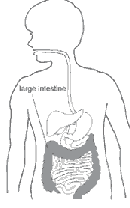 |
Why does HD cause constipation?
Normally, muscles in the intestine push stool to the anus, where stool leaves the body. Special nerve cells in the intestine, called ganglion cells, make the muscles push. A person with HD does not have these nerve cells in the last part of the large intestine.
In a person with HD, the healthy muscles of the intestine push the stool until it reaches the part without the nerve cells. At this point, the stool stops moving. New stool then begins to stack up behind it.
Sometimes the ganglion cells are missing from the whole large intestine and even parts of the small intestine before it. When the diseased section reaches to or includes the small intestine, it is called long-segment disease. When the diseased section includes only part of the large intestine, it is called short-segment disease.
 |
What causes HD?
HD develops before a child is born. Normally, nerve cells grow in the baby’s intestine soon after the baby begins to grow in the womb. These nerve cells grow down from the top of the intestine all the way to the anus. With HD, the nerve cells stop growing before they reach the end.
No one knows why the nerve cells stop growing. But we do know that it’s not the mother’s fault. HD isn’t caused by anything the mother did while she was pregnant.
Some children with HD have other health problems, such as Down’s syndrome and other rare disorders.
|
If I have more children, will they have HD too? |
 |
|
Symptoms in Newborns |
 |
Symptoms in Young Children
Most children with HD have always had severe problems with constipation. Some also have more diarrhea than usual.
|
Children with HD might also have anemia, a shortage of red blood cells, because blood is lost in the stool. Also, many babies with HD grow and develop more slowly than they should. |
 |
How does the doctor find out if HD is the problem?
To find out if a person has HD, the doctor will do one or more tests:
• barium enema x ray
• manometry
• biopsy
Barium Enema X Ray
An x ray is a black-and-white picture of the inside of the body. The picture is taken with a special machine that uses a small amount of radiation. For a barium enema x ray, the doctor puts barium through the anus into the intestine before taking the picture. Barium is a liquid that makes the intestine show up better on the x ray.
In some cases, instead of barium another liquid, called Gastrografin, may be used. Gastrografin is also sometimes used in newborns to help remove a hard first stool. Gastrografin causes water to be pulled into the intestine, and the extra water softens the stool.
In places where the nerve cells are missing, the intestine looks too narrow. If a narrow large intestine shows on the x ray, the doctor knows HD might be the problem. More tests will help the doctor know for sure.
Other tests to diagnose HD are manometry and biopsy:
Manometry
The doctor inflates a small balloon inside the rectum. Normally, the anal muscle will relax. If it doesn’t, HD may be the problem. This test is most often done in older children and adults.
Biopsy
This is the most accurate test for HD. The doctor removes and looks at a tiny piece of the intestine under a microscope. If the nerve cells are missing, HD is the problem.
The doctor may do one or all of these tests. It depends on the child.
What is the treatment?
Pull-through Surgery
HD is treated with surgery. The surgery is called a pull-through operation. There are three common ways to do a pull-through, and they are called the Swenson, the Soave, and the Duhamel procedures. Each is done a little differently, but all involve taking out the part of the intestine that doesn’t work and connecting the healthy part that’s left to the anus. After pull-through surgery, the child has a working intestine.
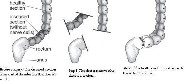 |
Colostonry and Ilostonry
Often, the pull-through can be done right after the diagnosis. However, children who have been very sick may first need surgery called an ostomy. This surgery helps the child get healthy before having the pull-through. Some doctors do an ostomy in every child before doing the pull-through.
In an ostomy, the doctor takes out the diseased part of the intestine. Then the doctor cuts a small hole in the baby’s abdomen. The hole is called a stoma. The doctor connects the top part of the intestine to the stoma. Stool leaves the body through the stoma while the bottom part of the intestine heals. Stool goes into a bag attached to the skin around the stoma. You will need to empty this bag several times a day.
 |
If the doctor removes the entire large intestine and connects the small intestine to the stoma, the surgery is called an ileostomy. If the doctor leaves part of the large intestine and connects that to the stoma, the surgery is called a colostomy.
Later, the doctor will do the pull-through. The doctor disconnects the intestine from the stoma and attaches it just above the anus. The stoma isn’t needed any more, so the doctor either sews it up during surgery or waits about 6 weeks to make sure that the pull-through worked.
What will my child’s life be like after surgery?
Ostomy
Most babies are more comfortable after having an ostomy because they can pass gas more easily and aren’t constipated anymore.
Older children will be more comfortable, too, but they may have some trouble getting used to an ostomy. They will need to learn how to take care of the stoma and how to change the bag that collects stool. They may be worried about being different from their friends. Most children can lead a normal life after surgery.
Nurses at the hospital can teach you and your child how to care for a stoma and can talk to you about your worries.
|
Adjusting After Pull-through Some children become constipated because 1 in 10 children with HD has difficulty moving stool through the part of the colon without nerve cells. |
 |
A mild laxative may also be helpful. Ask your doctor for suggestions.
Diet and Nutrition
One job of the large intestine is to collect the water and salts the body needs. Since your child’s intestine is shorter now, it absorbs less. Your child will need to drink more to make sure his body gets enough fluids.
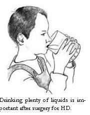 |
An infant who has long-segment disease requiring an ileostomy may need special tube feedings. The shortened intestine does not allow the bloodstream enough time to absorb nutrients from food before it is pushed out of the body as stool. Tube feedings that deliver nutrients can make up for what is lost.
Infection |
It can happen before or after surgery to treat Hirschsprung’s disease. Here are some of the signs to look for:
• fever
• swollen abdomen
• vomiting
• diarrhea
• bleeding from the rectum
• sluggishness
Call your doctor immediately if your child shows any of these signs. If the problem is enterocolitis, your child may be admitted to the hospital. In the hospital, an intravenous (I.V.) line may be needed to keep body fluids up and to deliver antibiotics to fight the infection. The large intestine will be rinsed regularly with a mild salt water solution until all remaining stool has been removed. The rinse may also contain antibiotics to kill bacteria.
When the child has recovered from the infection, the doctor may advise surgery. If the child has not had the pull-through surgery yet, the doctor may prepare for it by doing a colostomy or ileostomy before the child leaves the hospital. If the child has already had a pull-through operation, the doctor may correct the obstruction with surgery.
Enterocolitis can be life threatening, so watch for the signs and call your doctor immediately if they occur.
Sometimes HD affects most or all of the large intestine, plus some of the small intestine. Children with long-segment HD can be treated with pull-through surgery, but there is a risk of complications such as infection, diarrhea, and diaper rash afterward. Parents need to pay close attention to their child’s health. Also, since some, most, or all of the intestine is removed, drinking a lot of fluid is important.
Points to Remember
• HD is a disease of the large intestine.
• HD develops in children before they are born. It is not caused by anything the mother did while pregnant.
• Symptoms of HD include
|
|
delayed first bowel movement in newborns |
|
|
swollen abdomen and vomiting |
|
|
constipation since birth |
|
|
slow growth and development |
|
|
anemia |
• Children with HD may get an infection, called enterocolitis, which can cause fever and diarrhea.
• HD is a serious disease that needs to be treated right away. HD is treated with pull-through
surgery or, sometimes, ostomy.
• After treatment, most children with HD lead normal lives.
Acknowledgments
The National Digestive Diseases Information Clearinghouse would like to thank the following individuals who provided editorial guidance or facilitated field-testing for this publication.
Paul Hyman, M.D.
University of Kansas Medical Center
Kansas City, KS
Jacob C. Langer, M.D.
The Hospital for Sick Children
Toronto, Ontario, Canada
Andrea M. Anastas
International Foundation for Functional Gastrointestinal Disorders, Inc. (IFFGD) Pediatric
North Andover, MA
Kimberly Robinstein
Hirschsprung’s & Motility Disorders Support Network, The Guardian Society
Land O Lakes, FL
v Clostridium Difficile Infection
Clostridium difficile, or C. difficile (a gram-positive anaerobic bacterium), is now recognized as the major causative agent of colitis (inflammation of the colon) and diarrhea that may occur following antibiotic intake. C. difficile infection represents one of the most common hospital (nosocomial) infections around the world. In the United States alone, it causes approximately three million cases of diarrhea and colitis per year. This bacterium is primarily acquired in hospitals and chronic care facilities following antibiotic therapy covering a wide variety of bacteria (broad-spectrum) and is the most frequent cause of outbreaks of diarrhea in hospitalized patients. One of the main characteristics of C. difficile-associated colitis is severe inflammation in the colonic tissue (mucosa) associated with destruction of cells of the colon (colonocytes).
The disease involves, initially, alterations of the beneficial bacteria, which are normally found in the colon, by antibiotic therapy. The alterations lead to colonization by C. difficile when this bacterium or its spores are present in the environment. In hospitals or nursing home facilities where C. difficile is prevalent and patients frequently receive antibiotics, C. difficile infection is very common. In contrast, individuals treated with antibiotics as outpatients have a much smaller risk of developing C. difficile infection. Laboratory studies show that when C. difficile colonize the gut, they release two potent toxins, toxin A and toxin B, which bind to certain receptors in the lining of the colon and ultimately cause diarrhea and inflammation of the large intestine, or colon (colitis). Thus, the toxins are involved in the pathogenesis, or development of the disease.
Transmission factors - An important characteristic of C. difficile-associated diarrhea and colitis is its high prevalence among hospitalized patients. Thus, C. difficile contributes significantly to hospital length of stay, and may be associated in some elderly adults with chronic diarrhea, and occasionally other serious or potentially life-threatening consequences. One study demonstrated that 20% of patients admitted to a hospital for various reasons were either positive for C. difficile on admission or acquired the microorganism during hospitalization. Interestingly, only one-third of these patients developed diarrhea while the remainder were asymptomatic carriers serving as a reservoir of C. difficile infection. The organism and its spores were also demonstrated in the hospital environment, including toilets, telephones, stethoscopes, and hands of healthcare personnel.
While patient-to-patient spread and environmental contamination can be some of the reasons of cross-infection in C. difficile-associated diarrhea and colitis, antibiotic therapy is the major risk factor for this disease. Thus, antibiotic use only when necessary is the most effective measure of preventing C. difficile infection.
Clinical features - A wide range of conditions is associated with C. difficile infection. Most cases develop 4 to 9 days after the beginning of antibiotic intake. It should be noted, however, that some patients develop diarrhea after antibiotics are discontinued and this may lead to diagnostic confusion. Although nearly all antibiotics have been implicated with the disease, the commonest antibiotics associated with C. difficile infection are
ampicillin, amoxicillin, cephalosporins, and clindamycin.
The most common presentation is either mild colitis, or simple diarrhea that is watery and contains mucus but not blood. Examination by sigmoidoscopy usually reveals normal colonic tissue. General symptoms are commonly absent and diarrhea usually stops when antibiotics are discontinued. C. difficile can also cause nonspecific colitis quite reminiscent of other intestinal bacterial infections such as Shigella or Campylobacter. This is a more serious illness than simple antibiotic-associated diarrhea; patients experience watery diarrhea 10 to 20 times a day and lower, crampy abdominal pain. Low-grade fever, dehydration, and monospecific colitis are common manifestations.
Pseudomembranous colitis represents the characteristic manifestation of full-blown C. difficile-associated colitis. Sigmoidoscopic examination reveals the presence of characteristic plaque-like pseudomembranes, scattered over the colonic tissue. The presence of these plaques is a distinctive indicator of C. difficile infection in patients with diarrhea following antibiotic treatment.
The most serious manifestation of C. difficile infection, fulminant colitis (severe sudden inflammation of the colon), is frequently associated with very serious complications. This can be a life-threatening form of C. difficile infection and occurs in 3% of patients; most are elderly and debilitated from other diseases. Patients with this form of the disease experience severe lower abdominal pain, diarrhea, high fever with chills, and rapid heart beat. Timely treatment of fulminant colitis is essential; this condition can be life threatening.
C. difficile infection in patients with other intestinal diseases - It is well documented that C. difficile may complicate the course of ulcerative colitis or Crohn’s disease and it is responsible for 4 to 12% of diarrhea in AIDS patients. In this case, patients develop the typical symptoms of C. difficile colitis, including diarrhea, abdominal pain, and fever reminiscent of exacerbation of inflammatory bowel disease. The reason for this complication is not entirely clear. It may be that the frequent hospitalizations and exposure to antibiotics of patients with inflammatory bowel disease or AIDS places them at increased risk for the infection. So far there is no evidence to indicate that C. difficile can complicate the symptoms associated with irritable bowel syndrome (IBS).
Laboratory diagnosis - The laboratory diagnosis of C. difficile infection is primarily related to the demonstration of C. difficile toxins in the stool of suspected patients. The detection of C. difficile toxins in the stool can be made by a laboratory test (cytotoxicity assay) where the toxins can be easily observed in the microscope. This tissue culture assay is considered the gold standard because of its high sensitivity and specificity. Since there is no correlation between levels of C. difficile toxins in the stool and severity of the disease, the results are reported simply as “positive” or “negative.” However, time is a drawback of this assay since it requires 24 to 48 hours to read the results.
Over the past few years several rapid tests that take just a few hours, and which do not require specialized personnel to run, have been developed (immuno-enzymatic assays) for the detection of C. difficile toxins in the stool. These tests are commercially available in the form of diagnostic kits. Although they are relatively less sensitive and demonstrate lower specificity compared to the laboratory tests, they are very useful not only in the every day practice when specialized personnel is not available, but also in emergency situations and in rapid screening of patients during spreading of the disease in hospitals.
Therapy - Therapy of C. difficile is directed against eradication of the microorganism from the colonic microflora. No therapy is required for asymptomatic carriers. In noncomplicated patients with mild diarrhea, no fever, and modest lower abdominal pain, discontinuation of antibiotics (if possible) is often enough to alleviate symptoms and stop diarrhea. When severe diarrhea is present and in cases of established colitis, the patients should receive the antibiotics, metronidazole or vancomycin, for 10 to 14 days. Several clinical trials have shown that these antibiotics are equally effective in cases of mild to moderate C. difficile infection and more than 95% of patients respond very well to this treatment. Diarrhea following treatment with either vancomycin or metronidazole is expected to improve after 1 to 4 days with complete resolution within 2 weeks. However, some patients do not respond despite aggressive medical therapy and require surgical intervention.
Therapy for relapsing C. difficile infection - Although C. difficile infection usually responds well to treatment with metronidazole or vancomycin, approximately 15 to 20% of patients will experience reappearance of diarrhea and other symptoms weeks or even months after initial therapy has been discontinued. The usual therapy for relapse is to repeat the 10 to 14 day course of either metronidazole or vancomycin and this is successful in most patients. However, a subset of patients continues to relapse whenever antibiotics are discontinued and this represents a therapeutic challenge. Some authorities recommend switching to the alternative antibiotic from the one used initially. A variety of other therapies have also been described for relapsing disease. It is hoped that development of vaccines against C. difficile toxins may someday control the problem of C. difficile infection in hospitals.
by: Charalabos Pothoulakis, M.D., Division of Gastroenterology, Beth Israel Deaconess Medical Center, Harvard Medical School, Boston, MA.
@2004 IFFGD. This article is reprinted with permission from the International Foundation for Functional Gastrointestinal Disorders (IFFGD). If you would like more information on gastroinstestinal disorders contact IFFGD, 1-888-964-2001 (toll free) or visit their website at www.iffgd.org.
For more information
American Academy of Family Physicians
P.O. Box 11210
Shawnee Mission, KS 66207-1210
Phone: 1-800-274-2237
Email: fp@aafp.org
Internet: www.aafp.org
American College of Surgeons
633 North Saint Clair Street
Chicago, IL 60611-3211
Phone: (312) 202-5000
Fax: (312) 202-5001
Email: postmaster@facs.org
Internet: www.facs.org
American Gastroenterological Association
4930 Del Ray Avenue
Bethesda, MD 20814
Phone: (301) 654-2055
Fax: (301) 654-5920
Email: info@gastro.org or webmaster@gastro.org
Internet: www.gastro.org
American Liver Foundation (ALF)
75 Maiden Lane, Suite 603
New York, NY 10038-4810
Phone: 1-800-GO-LIVER (465-4837),
1-888-4HEP-USA (443-7872),
or (212) 668-1000
Fax: (212) 483-8179
Email: info@liverfoundation.org
Internet: www.liverfoundation.org
American Society of Colon and Rectal Surgeons (ASCRS)
85 West Algonquin Road
Suite 550
Arlington Heights, IL 60005
Phone: (847) 290-9184
Fax: (847) 290-9203
Email: ascrs@fascrs.org
Internet: www.fascrs.org
American Porphyria Foundation
P.O. Box 22712
Houston, TX 77227
Phone: (713) 266-9617
Email: porphyrus@aol.com
Internet: www.porphyriafoundation.com
Cyclic Vomiting Syndrome Association
3585 Cedar Hill Road, NW.
Canal Winchester, OH 43110
Phone: (614) 837-2586
Fax: (614) 837-2586
Email: waitesd@cvsaonline.org
Internet: www.cvsaonline.org
International Foundation for Functional Gastrointestinal Disorders, Inc. (IFFGD)
P.O. Box 170864
Milwaukee, WI 53217-8076
Phone: 1-888-964-2001 or (414) 964-1799
Fax: (414) 964-7176
Email: iffgd@iffgd.org
Internet: www.iffgd.org
National Library of Medicine—MEDLINEplus
Internet: www.nlm.nih.gov/medlineplus
National Organization for Rare Disorders Inc. (NORD)
55 Kenosia Avenue
P.O. Box 1968
Danbury, CT 06813-1968
Phone: 1-800-999-6673 or (203) 744-0100
Fax: (203) 798-2291
Email: orphan@rarediseases.org
Internet: www.rarediseases.org
Pull-thru Network
2312 Savoy Street
Hoover, AL 35226
Phone: (205) 978-2930
Email: info@pullthrough.org
Internet: www.pullthrough.org
Wilson’s Disease Association
4 Navaho Drive
Brookfield, CT 06804
Phone: 1-800-399-0266 or (203) 775-9666
Email: hasellner@worldnet.att.net
Internet: www.wilsonsdisease.org