v Terminology
n Groups of Teeth
Teeth, as they exist in the mouth, can be placed into any of three broad groupings, the maxillary or mandibular, right or left, anteriors or posteriors. These groupings apply to both the natural dentition and to artificial teeth:
¨ Maxillary or Mandibular
A person has two jaws, a maxillary (upper) and a mandibular (lower). The teeth in these jaws are called either maxillary or mandibular teeth. The combination of natural teeth and supporting alveolar bone that is found in an upper or a lower jaw is called a dental arch . When natural teeth are extracted, the healed alveolar process is called the residual ridge. Artificial teeth sit over residual ridges so they coincide with the original arch form.
¨ Right or Left
If we split the two dental arches down the midline from front to back, the arches can be divided into upper and lower right sections and upper and lower left sections. Since one of these sections represents one-fourth of the upper and lower arches taken together, the section is called a quadrant (Figure 2-1). If a tooth is located to the left of the midline in the upper arch, the tooth is part of the maxillary left quadrant (etc.).
¨ Anteriors or Posteriors (Figure 2-2)
Teeth can also be classified as anteriors (incisors and cuspids) or posteriors (bicuspids and molars). A complete adult natural dentition has 32 teeth; each arch contains 16. The teeth in an arch are composed of 6 anteriors (cuspid to cuspid) and 10 posteriors (all teeth distal to the cuspids). There are 3 anteriors and 5 posteriors in a quadrant.
Figure 2-1. Mandibular Right Quadrant
![]()
Anterior Posterior Teeth
Figure 2-2. Anterior and Posterior Teeth
NOTE: Complete dentures for the upper and lower arches usually consist of 28 teeth. The 4 third molars are not used.
n Names of Teeth (Figure 2-3)
¨ Anteriors
|
Central and Lateral Incisors. In each quadrant, the two teeth nearest the midline of the dental arches are called incisors. The first incisor on either side of the midline is called a central incisor. The second incisor from the midline of either arch is called a lateral incisor. The word incisor describes their functions of incising or cutting food. | |
|
Cuspids . A cuspid is so named because its cutting edge is a single, pointed elevation or cusp. Cuspids are sometimes called canines. They are used to tear food. Each dental arch has two cuspids. |
¨ Posteriors
The two biscupids in any given quadrant
|
 |
||||
n Number Substitutes for Names of Teeth (Figure 2-4)
Formal descriptions like "maxillary right molar" and "mandibular left lateral incisor" can be time- consuming when many people must be examined in a short time, and too lengthy when space on forms is limited. Dentists often use numerical shorthand as a substitute for complete, formal tooth names. The full complement of natural teeth is numbered #1 through #32. Numbers 1 through #16 are in the maxillary arch; the upper right third molar is #1, the upper right second molar is #2, and as you proceed in consecutive order around the maxillary arch to the upper left third molar, you end with #16. Numbers 17 through #32 are in the mandibular arch; the lower left third molar is #17, the lower left second molar is #18, and as you proceed around the mandibular arch to the lower right third molar you end with #32.

Figure 2-4. Number Substitutes for Names of Teeth
|
1. Right maxillary third molar. 2. Right maxillary second molar. 3. Right maxillary first molar. 4. Right maxillary second bicuspid. 5. Right maxillary first bicuspid. 6. Right maxillary cuspid 7. Right maxillary lateral incisor. 8. Right maxillary central incisor. 9. Left maxillary central incisor. 10. Left maxillary lateral incisor. 11. Left maxillary cuspid. 12. Left maxillary first bicuspid. 13. Left maxillary second bicuspid. 14. Left maxillary first molar. 15. Left maxillary second molar. 16. Left maxillary third molar. |
17. Left mandibular third molar. 18. Left mandibular second molar. 19. Left mandibular first molar. 20. Left mandibular second bicuspid. 21. Left mandibular first bicuspid. 22. Left mandibular cuspid. 23. Left mandibular lateral incisor. 24. Left mandibular central incisor. 25. Right mandibular central incisor. 26. Right mandibu!ar lateral incisor. 27. Right mandibular cuspid. 28. Right mandibular first bicuspid. 29. Right mandibular second bicuspid. 30. Right mandibular first molar. 31. Right mandibular second molar. 32. Right mandibular third molar. |
 |
n Structure of the Teeth and Supporting Tissues (Figure 2-5)
¨ Teeth
A tooth is divided into two parts, the crown and the root. The anatomical crown is the part of the tooth covered with enamel. The root of a tooth is embedded in alveolar bone and covered with cementum.
NOTE: In young people, areas of the anatomical crown are frequently buried in gingival tissue. As a person gets older it becomes common for a tooth’s enamel to be completely exposed above the gingiva and to have root surface showing. The term clinical crown is applied to the part of the tooth that is visible above the gingiva to include root surface.
The bulk of a tooth is composed of a bone-like substance called dentin which is covered by enamel to form the crown and cementum to form the root. The line of division between the crown and root is called the cervical line or cemento enamel junction. The dividing line is found in a somewhat constricted region on the tooth’s surface called the cervix or neck. The tip of the root is known as the apex.
The tooth contains an aggregate of blood vessels, nerves, and cellular connective tissue called the dental pulp. The dental pulp is housed within a pulp chamber and root canal of a tooth. Anterior teeth ordinarily have one root canal; multiple canals occur in posterior teeth. The nerves and blood vessels enter and leave the tooth through an opening called the apical foramen at or near the apex of the root.
 |
¨ Supporting Structures of the Teeth
The supporting tissues of the teeth are collectively called the periodontium. The periodontium consists of the alveolar process of the maxillae and the mandible, the periodontal ligament, the cementum of the tooth, and the gingiva.
|
|
Alveolar Process. The alveolar process is the portion of the maxillae or mandible in which the roots of the teeth are embedded and by which tooth roots are supported. An alveolar process consists of three kinds of bone. They are the outer cortical plate, lamina dura, and spongy bone. The outer cortical plate is a compact layer of bone on the bone’s surface. The lamina dura is a thin, dense layer of bone that lines tooth sockets. The lamina aura is a specialized continuation of the cortical plate. Spongy bone is the less dense, cancellous bone representing the alveolar process’ central mass. |
|
|
Periodontal Ligament. The periodontal ligament is a thin, fibrous ligament connecting a tooth to the lamina aura of the bony socket. Normally, teeth do not contact the bone directly; a tooth is suspended in its socket by the fibers of the ligament. This arrangement allows each tooth limited individual movement. The fibers act as shock absorbers to cushion the force of chewing impacts. |
|
|
Cementum. The cementum is the only tissue considered as both a basic part of the tooth and a component of the periodontium. It is a thin, calcified layer of tissue that completely covers the dentin of a tooth’s root. Cementum is formed during the development of the root. It functions as an area of attachment for periodontal ligament fibers. |
|
|
Gingiva (Figure 2-6). The gingiva is the specialized mucous membrane covering the alveolar processes and encircling the necks of the teeth. It aids in the support of the teeth, and protects the alveolar process and periodontal ligament from bacterial invasion. Healthy gingiva is pale pink, firm, and resilient. It is divided into two types, free and attached gingiva. |
Free gingiva is "free" to the extent that it can be displaced; it is not tightly bound to anything underneath it. Free gingiva extends from the gingival crest to the bottom of the gingival sulcus. At the bottom of the sulcus, an epithelial attachment joins the free gingiva to the tooth surface. The interdental papilla is the portion of the free gingiva that fills the proximal space below the contact areas of adjacent teeth. It helps prevent food from packing between the teeth.
Attached gingiva covers the labial cortical plate of the alveolar process. It is firmly fixed to underlying bone.
n Crown Morphology (Contours)
As you study anatomy, you will discover there are almost no perfectly flat or perfectly straight surfaces; most surfaces are curved. The contour of a crown is a combination of convex and concave curves. A convex surface is one which is curved outward; a concave is curved inward.
¨ Tooth Surfaces (Figure 2-7)
|
| Proximal. A tooth has two proximal surfaces, one that is oriented toward the midline of the dental arch and another that is oriented away from the midline of the arch. |
- Mesial. The mesial is the proximal surface closest to the midline of the arch.
- Distal . The distal is the proximal surface oriented away from the midline of the arch.
|
| Facial. The facial is the surface of a tooth that "faces" toward the lips or cheeks. When there is a requirement to be more specific, terms like labial and buccal are used: |
- Labial . The labial is the surface of an anterior tooth that faces toward the lips.- Buccal. The buccal is the surface of a posterior tooth that faces toward the cheek.
|
|
Lingual .The surface of a tooth facing toward the tongue is called the lingual. |
|
|
Incisal. The cutting edge of an anterior tooth. |
|
|
Occlusal. The chewing surface of a posterior tooth. |
|
Figure 2-7. Tooth Surface |
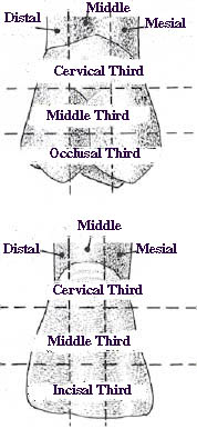
Figure 2-9. Anterior and Posterior Crown Divisions |
Figure 2-8. Long Axis |
|
 |
- Anterior Tooth Line Angles
1. Mesiolabial
2. Mesiolingual
3. Distolabial
4. Distolingual
5. Labioincisal
6. Linguoincisal
7. Mesioincisal
8. Distoincisal
- Posterior Tooth Line Angles
1. Mesiobuccal
2. Mesiolingual
3. Distobuccal
4. Distolingual
5. Bucco occlusal
6. Linguo occlusal
7. Disto occlusal
8. Mesio occlusal
|
| Point Angle |
- Anterior Tooth Point Angles
1. Mesiolabioincisal
2. Mesiolinguoincisal
3. Distolabioincisal
4. Distolinguoincisal
- Posterior Tooth Point Angles
1. Mesiobucco-occlusal
2. Mesiolinguo-occlusal
3. Distobucco-occlusal
4. Distolinguo-occlusal
¨ Distinctive Crown Convexities and Concavities
|
| Convexities |
|
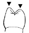
Lobe (Figure 2-10) . A lobe is one of the primary anatomical divisions of a crown; all teeth develop from either four or five lobes (for example, a central incisor forms from four lobes while first molars develop from five lobes.) Lobes are usually separated by readily identifiable developmental grooves. Mamelons (Figure 2-11). Mamelons are small, rounded projections of enamel from the incisal edges of newly erupted anterior teeth. The projections wear away soon after eruption. Cingulum (Figure 2-12). A cingulum is found on the lingual aspect of an anterior tooth. It is a convex mount of enamel localized to the cervical one-third of the crown. |
 |
| Figure 2-11. Mamelons | |
 |
|
| Figure 2-12. Cingulum |
|
Cusp (Figure 2-13). A cusp is a pointed or rounded elevation of enamel found on cuspids and on the chewing surfaces of bicuspids and molars. Cuspids have one cusp that represents the tooth’s cutting edge. Maxillary bicuspids and the mandibular first bicuspids have two cusps, one buccal and one lingual. The mandibular second bicuspid normally has three cusps, one buccal and two lingual. The lingual cusps are subdivided into a mesiolingual and a distolingual. |
 |
| Figure 2-13. Cusp |
All maxillary molars have four cusps, two buccal and two lingual. The two buccal cusps are subdivided into a mesiobuccal and a distobuccal. The two lingual cusps are subdivided into a mesiolingual and a distolingual. (Once in a while, the mesiolingual cusp of a maxillary first molar carries an underdeveloped, rudimentary cusp called the cusp of Carabelli.)
The mandibular first molar has five cusps, three buccal and two lingual. From anterior to posterior, the three buccal cusps are subdivided into a mesiobuccal, a distobuccal, and a distal. The two lingual cusps are divided into a mesiolingual and a distolingual.
The mandibular second molar has four cusps called the mesiobuccal, distobuccal, mesiolingual, and distolingual.
Ridge. Any linear elevation found on the surface of a tooth.
|
Marginal Ridge (Figure 2-14). A marginal ridge is a linear, rounded border of enamel that forms the mesial and distal margins of anterior teeth as viewed from the lingual, and the mesial and distal borders of occlusal surfaces on posterior teeth. Lingual Ridge (Figure 2-15). The ridge of enamel that extends from the cingulum to the cusp tip on the lingual surface of most cuspids is called the lingual ridge. Cusp Ridge (Figure 2-16). Each cusp has four cusp ridges radiating from its tip. They are named according to the direction they take away from the cusp tip (for example, mesial, distal, facial, or lingual). |
 |
| Figure 2-14. Marginal Ridge | |
 |
|
| Figure 2.15. Lingual Ridge |
 |
Triangular Ridge (Figure 2-17). The occlusal surface of a cusp is composed of a mesial and a distal incline. These two inclines meet to form a triangular ridge of enamel that descends from the tip of the cusp to the central portion of the occlusal surface. A triangular ridge is either a facial or a lingual cusp ridge, depending on where the cusp is located. Cusps are described in some mouths as being "pointy" and in others as being "flat" or "blunt." Most "pointy" posterior teeth have high cusp angle values (Figure 2-18). A cusp angle is the angle that a triangular ridge makes with a plane perpendicular to the long axis of the tooth.

Figure 2-17. Triangular Ridge |
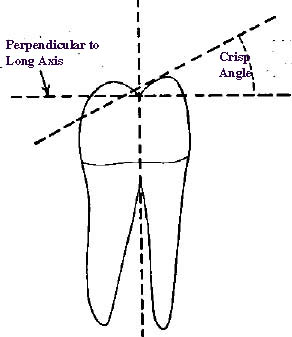
Figure 2-18. Cusp Angle |
|
Tr ansverse Ridge (Figure 2-19). A transverse ridge is the union of a buccal and lingual triangular ridge that crosses the surface of a posterior tooth transversely (roughly 90 degrees to both the buccal and lingual tooth surfaces). Oblique Ridge (Figure 2-20). The only tooth on which an oblique ridge is found is the maxillary molar. An oblique ridge consists of a union between the triangular ridge of the distobuccal cusp and the distal cusp ridge of the mesiolingual cusp. Cusp Inclines. A cusp incline or inclined plane is the sloping area found between two cusp ridges. To name an incline, you must combine the names of the cusp ridges that define a large part of its borders, for example, the distolingual incline of the buccal cusp of a maxillary first bicuspid (Figure 2-16). • Crown Concavities Fossae Lingual Fossa ( Figure 2-21). The lingual fossa is an irregular, rounded concavity bound by the mesial marginal ridge, distal marginal ridge, cingulum, and incisal edge of the lingual surface of an incisor tooth. Lingual fossae are also found on both sides of the lingual ridge of a cuspid tooth. Triangular Fossa (Figure 2-22). Triangular fossae are located adjacent to marginal ridges on the occlusal surfaces of posterior teeth. There are two kinds of triangular fossae, a mesial and a distal. |
Figure 2-19. Tranverse Ridge |
Figure 2-20. Obligue Ridge |
|
Figure 2-21. Lingual Fossa |
|

Figure 2-22. Triangular Fossa |
|
|
Central Fossa (Figure 2-23). A central fossa is a centrally located depression or concavity found on the occlusal surface of molars and mandibular second bicuspids. The other bicuspids have mesial and distal triangular fossae, but do not have a central fossa. Sulcus (Figure 2-24). A sulcus is an elongated valley or depression in the surface of a tooth formed by the inclines of adjacent cusps or ridges. As an example, a central sulcus is a major linear depression that traverses the occlusal surface of a posterior tooth from mesial triangular fossa to distal triangular fossa. Developmental grooves are found in the bottoms of sulci. Developmental Groove (Figure 2-25). A developmental groove is the junction line between the inclined walls of a sulcus. Developmental grooves represent lines of union between lobes of the crown during its formation. These grooves appear on labial, occlusal, buccal, and lingual surfaces, and are least apparent on the labial aspect of anteriors. Supplemental Groove (Figure 2-26). A minor, auxiliary groove that branches off from a much more prominent developmental groove. Supplemental grooves do not represent the junction of primary tooth parts. Fissure (Figure 2-27). A linear fault that sometimes occurs in a developmental groove. A fissure represents a lack of union between the inclined walls of a sulcus. Pit . A pit is a small, pinpoint fault on the surface of a tooth; a pit is usually found at the end of a developmental groove or at a place where two fissures intersect. |
Maxillary Molar |
Mandibular Molar Figure 2-23. Central Fossa |
|
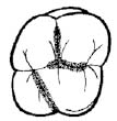
Figure 2-24. Sulcus |
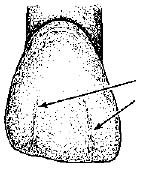 |
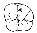
Figure 2-26. Supplemental Groove |

Figure 2-25. Developmental Grooves |

Figure 2-27. Fissure |
|
Maxillary Central Incisor (Facial View) 1. Lobe 2. Developmental Groove 3. Cemento–Enamel Junction 4. Incisal Edge |
 |
| Figure 2.28. Maxillary Central Incisor (Facial View) | |
Application. Figures 2-28 through 2-33 show specific convexities and depressions on anterior and posterior teeth. You should be able to name the coronal features of teeth after you study these figures closely.
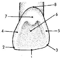
Figure 2-29. Maxillary Central Incisor (Lingual View)
1. Incisal Edge 5. Distal Marginal Ridge
2. Mesio-Inciso Angle 6. Lingual Fossa
3. Disto-Inciso Angle 7. Cingulum
4. Mesial Marginal Ridge 8. Cemento-Enamel Junction
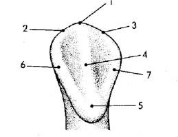
Mandibular Cuspid (Lingual View)
1. Cusp Tip 5. Cingulum
2. Mesial Cusp Ridge 6. Mesial Marginal Ridge
3. Distal Cusp Ridge 7. Distal Marginal Ridge
4. Lingual Ridge
Figure 2-30. Mandibular Cuspid (Lingual View)
Maxillary Second Bicuspid (Occlusal View)
|
1. Buccal Cusp 2. Lingual Cusp 3. Central Sulcus (Developmental Groove) 4. Supplemental Groove 5. Mesial Marginal Ridge 6. Distal Marginal Ridge |
7. Mesial Triangular Fossa 8. Distal Triangular Fossa 9. Buccal Triangular Ridge (Crest) 10. Lingual Triangular Ridge (Mesial Incline) 11. Transverse Ridge 12. Mesial Cusp Ridge 13. Distal Cusp Ridge |
Figure 2.31.Maxillary Second Bicuspid (Occlusal View)
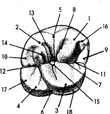
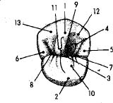
Maxillary First Molar (Occlusal View)
|
1. Mesio-BuccalCusp 2. Disto-Buccal Cusp 3. Mesio-Lingual Cusp 4. Disto-Lingual Cusp 5. Buccal Developmental Groove 6. Lingual Developmental Groove 7. Central Sulcus (Developmental Groove) 8. Supplemental Groove 9. Mesial Marginal Ridge |
10. Distal Marginal Ridge 11. Mesial Triangular Fossa 12. Distal Triangular Fossa 13. Central Fossa 14. Disto Buccal Triangular Ridge (Crest) 15. Oblique Ridge 16. Mesial CuspRidge 17. Distal Cusp Ridge 18. Cusp of Carabelli |
Figure 2.32 Maxillary First Molar (Occlusal View)
Mandibular First Molar (Occlusal View)
|
1. Mesio-Buccal Cusp 2. Disto-Buccal Cusp 3. Distal Cusp 4. Mesio-Lingual Cusp 5. Disto-Lingual Cusp 6. Buccal (Developmental Groove) 7. Central Sulcus 8. Supplemental Groove 9. Mesial Marginal Ridge |
10. Distal Marginal Ridge 11. Mesial Triangular Fossa 12. Distal Triangular Fossa 13. Central Fossa 14. Disto-Buccal Triangular Ridge (Distal Incline) 15. Disto-Lingual Triangular Ridge (Crest) 16. Tranverse Mesial 17. Mesial Cusp Ridge 18. Distal Cusp Ridge |
Figure 2.33. Mandibular First Molar (Occlusal View)
|
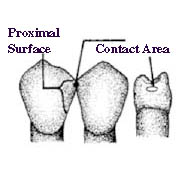
Figure 2.34. Contact Area |

Round |
|
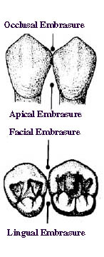
Figure 2-35. Embrasures |
Rectangular |
Trapezoid |
|
Rhomboid Figure 2.36. Occlusal Surface Outlines of Posterior Teeth |
n Proximal Surface Contact Characteristics
¨ Contact Points or Areas (Figure 2-34)
Teeth make contact with one another at points or areas on the greatest contour of their proximal surfaces. The places where adjacent teeth make point contact are called contact points. Contact points become wider and flatter in time from wear that occurs during functional movements (chewing) or parafunctional movements (grinding). A flattened contact point is called a contact area.
¨ Embrasure (Figure 2-35)
An embrasure is a space diverging from the contacting proximal surfaces of two adjacent teeth. There are four of these spaces or embrasures recognized. They are the facial, lingual, gingival, and occlusal or incisal (depends on whether they are posterior or anterior teeth). The gingival embrasure is located cervical to the contacting areas of adjacent teeth. A gingival embrasure has other names like cervical embrasure, apical embrasure, interproximal space, and septal space. Interdental palillae (gingival tissue) fill interproximal spaces to a greater or lesser extent.
n Occlusal Surface Outlines of Posterior Teeth (Figure 2-36)
¨ Circular Rounded in Outline
The occlusal surfaces of the lower bicuspids are circular in outline.
¨ Rectangular
The occlusal surfaces of the lower second molar and the upper bicuspids are often described as being rectangular or oblong in outline.
¨ Trapezoid
A trapezoid is a plain four-sided figure with two parallel sides. The occlusal surface of the lower first molar is said to be trapezoidal in outline.
¨ Rhomboidal
A rhomboid is shaped as an equilateral parallelogram having two opposing oblique angles. The occlusal surfaces of the upper molars are rhomboidal in outline.
v Descriptions of Individual Teeth
n Overview
In the following pages each tooth of the permanent dentition is described, except for the third molars, which are not reproduced in artificial teeth. In each instance, the tooth from the right side of the mouth is illustrated. The teeth are described as they usually look, but it should be mentioned that teeth vary considerably from one person to another and that certain teeth in the dentition tend to vary more than others. Included in the illustrations of the bicuspids and the molars are drawings that show angles that can be carved in reproducing the occlusal surfaces of these teeth. The broken lines shown in the illustrations of the facial and lingual surfaces of the teeth indicate proper food deflection contours.
The drawings in this section were adapted from those appearing in the Ney Crown and Bridge Manual, J. M. Ney Co., Hartford, Conn.
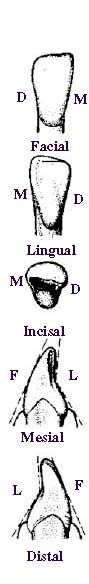
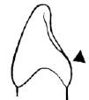
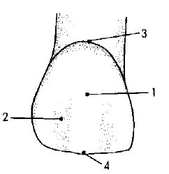
n Maxillary Central Incisor
The maxillary central incisor (Figure 2-37) is the tooth nearest the median line in the maxillary arch.
¨ The facial surface is broad and resembles a thumbnail in outline. The right maxillary central incisor may be distinguished from the left maxillary central incisor because the distoincisal angle is more rounded than the mesioincisal angle, and the incisal edge slopes slightly gingivally in a mesiodistal direction. The facial surface is convex both mesiodistally and incisocervically. Three distinct lobes may be seen in the incisal portion; they are separated by two developmental grooves.
¨ The lingual surface appears slightly smaller than the facial surface and the cervical portion is narrower. The large lingual fossa is bounded by prominent mesial and distal marginal ridges. There is a cingulum in the cervical portion, and there may be a pit in conjunction with the cingulum.
¨ Viewed on end, the incisal edge appears nearly straight. Most of the wear is on the lingual portion of the edge, so the edge becomes beveled lingually. The cingulum lies more to the distal side of the tooth than to the mesial side.
¨ The mesial surface looks like a wedge. The apex of the wedge is at the incisal edge of the tooth. The facial outline is slightly convex. The lingual outline is slightly concave from the incisal edge to the cingulum and convex from the cingulum to the cervical margin.
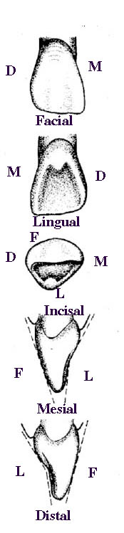
Figure 2-37. Maxillary Central Incisor |
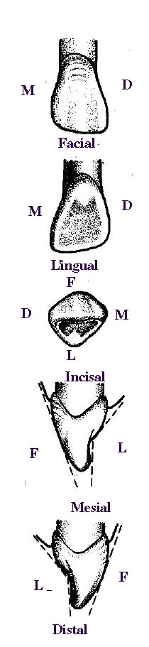
Figure 2-38. Maxillary Lateral Incisor |
¨ The distal surface closely resembles the mesial surface. The lingual outline is more concave in the incisal portion than it is on the mesial surface.
n Maxillary Lateral Incisor
The maxillary lateral incisor (Figure 2-38) is the second tooth from the median line in the maxillary arch. It resembles the central incisor but is smaller in all dimensions.
¨ The facial surface is narrower and shorter than that of the central incisor. The distoincisal angle is more rounded than the mesioincisal angle. The distal portion of the incisal ridge slopes upward toward the distoincisal angle. The facial surface is convex.
¨ The lingual surface resembles the facial surface in peripheral outline except that the cervical portion is narrower. The features of this surface vary considerably from one individual to another. Proportionally, the lingual surface characteristics of a lateral incisor are more marked than similar features on a central incisor.
¨ Viewed on end, the incisal edge appears nearly straight. The cingulum lies slightly to the distal side of the tooth.
¨ The mesial surface, like that of the central incisor, is wedge-shaped. The apex of the wedge is at the incisal edge. The incisal edge lies somewhat further lingually than it does in the central incisor.
¨ The distal surface resembles the mesial surface, but the facial outline is more convex and the incisal portion of the lingual outline is more concave.
n Maxillary Cuspid
The maxillary cuspid (Figure 2-39) is the third tooth from the median line in the maxillary arch. It is located at the corner of the arch, and its long root is embedded in the canine (cuspid) eminence. The maxillary cuspid is usually the longest tooth in either jaw. Since it resembles a dog’s tooth, it is sometimes called the canine.
¨ The incisal portion of the facial surface is much broader than the cervical portion. The mesial and distal cusp ridges of the incisal edge slope downward toward the center to meet at the tip of the cusp. The distal slope is longer than the mesial slope. The facial surface is convex. It is divided into mesial and distal surfaces by the facial ridge. The ridge extends from the tip of the cusp to the point of greatest convexity. The mesiofacial surface of the cuspid falls on the curve of the arch formed by the anterior teeth. The distofacial surface conforms to the buccal alignment of posterior teeth.
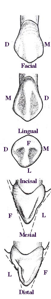
Figure 2-39. Maxillary Cuspid |
¨ The lingual surface resembles the facial surface in outline, but the cervical portion is narrower. The mesial and distal marginal ridges prominent, and a strong lingual ridge runs from the tip of the cusp to the cingulum. The maxillary cuspid has the largest cingulum of all the anterior teeth. ¨ Viewed on end, the incisal edge is slightly curved. The lingual portion of the tooth appears rugged, with the ridges and grooves being very well defined. ¨ The mesial surface is roughly triangular. From this aspect, the cuspid appears much thicker than the incisors. ¨ The distal surface is shaped very much like the mesial surface but is shorter because the distal portion of the incisal edge slopes further cervically than the mesial portion. n Maxillary First Bicuspid The maxillary first bicuspid (Figure 2-40) is the fourth tooth from the median line in the maxillary arch. It is the first posterior tooth. The bicuspids are sometimes called premolars because they are just in front of the molars: ¨ The facial surface resembles that of the cuspid in outline, but it is shorter occlusocervically and not quite as convex. The slopes of the mesial and distal cusp ridges are about equal in length. The facial ridge is prominent. ¨ The lingual surface is much smaller than the facial surface in all dimensions but is generally similar in outline. The lingual cusp is shorter than the facial cusp and is located mesial to the midline of the tooth. ¨ The occlusal surface is broader facially than lingually. There are two cusps, the facial cusp and the lingual cusp. The mesial and distal marginal ridges correspond to the marginal ridges of the anterior teeth. The proximal surfaces converge toward the lingual; the distal surface converging the most. |
|
The mesial fossa is distal to the mesial marginal ridge and the distal fossa is mesial to the distal marginal ridge. The facial and lingual triangular ridges extend from the tips of the cusps to the central groove. This groove ends at the mesial and distal pits. The mesial and distal marginal grooves arise from the mesial and distal pits and end on the mesial and distal surfaces, respectively. ¨ The mesial surface is roughly rectangular in outline. The facial and lingual outlines are convex. The mesial surface is generally convex except for a concave area on the facial portion of the surface above the cervical margin. The mesial marginal groove extends onto the mesial surface. ¨ The distal surface resembles the mesial surface but does not have the concave area above the cervical margin. n Maxillary Second Bicuspid The maxillary second bicuspid is the fifth tooth from the median line in the maxillary arch. It closely resembles the first bicuspid but is more rounded in outline (Figure 2-41). ¨ The lingual surface is only slightly shorter than the facial surface because the facial and lingual cusps are nearly equal in length. This surface is also slightly narrower than the facial surface. The lingual surface is smoothly convex in all directions, and its greatest convexity is in the cervical third. ¨ The occlusal surface has, in general, the same form and features as the occlusal surface of the first bicuspid. However, the facial and lingual portions are more nearly equal in size and the mesial and distal pits are closer together. ¨ The mesial surface is wider in the cervical portion than in the occlusal portion. The facial outline is slightly convex, except in the central portion. The lingual outline is convex. Both cusps appear more rounded that the cusps of the first bicuspid. ¨ The distal surface is slightly shorter than the mesial surface; however, it is about the same width. The facial and lingual outlines are convex. The surface is smoothly convex except at the distal marginal groove. n Maxillary First Molar The maxillary first molar (Figure 2-42) is the sixth tooth from the median line in the maxillary arch. It is the largest tooth in either arch. The maxillary and mandibular first molars are often called 6-year molars: ¨ The facial surface is roughly heart shaped in outline. The mesiofacial and distofacial cusps form the occlusal border and the facial groove divides the cusps. The surface is generally convex, except at this groove. The surface has three ridges. A ridge extends perpendicularly from the tip of each cusp and a third ridge extends horizontally in the cervical portion. ¨ The mesiolingual and distolingual cusps outline the occlusal border of the lingual surface. The mesiolingual cusp is the largest of the posterior teeth. Quite often this tooth has a fifth cusp, the cusp of Carabelli, which is on the lingual surface of the mesiolingual cusp. This cusp, when present, is shorter than the other cusps and does not form part of the occlusal surface. The lingual surface is generally convex, except at the distolingual groove. |
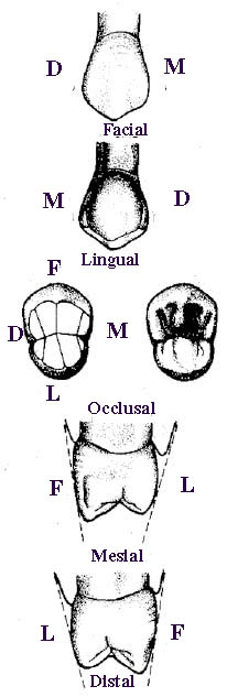
Figure 2-40. Maxillary First Bicuspid
Figure 2-41. Maxillary Second Bicuspid |
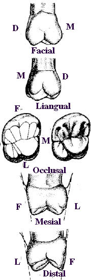
Figure 2.42. Maxillary First Molar
Figure 2.43. Maxillary Second Molar |
¨ The occlusal surface is roughly rhomboidal. The cusps are large and prominent, with broad surfaces broken up into rugged ridges and well-defined grooves. The mesiolingual cusp is the largest of the cusps. The distolingual groove separates it from the distolingual cusp. An oblique ridge connects the mesiolingual and distofacial cusps. It runs parallel to the distolingual groove. The facial groove runs from the central pit onto the facial surface. The mesial and distal pits lie near the mesial and distal marginal ridges, respectively. ¨ The mesial marginal groove which starts at the mesial pit notches the occlusal border of the mesial surface. A double convexity marks the lingual margin if the cusp of Carabeli is present. ¨ The distal marginal groove, which starts at the distal pit notches the occlusal border of the distal surface. n Maxillary Second Molar The maxillary second molar (Figure 2-43) is the seventh tooth from the median line in the maxillary arch. It is quite similar to the first molar; however, it is smaller. The tooth is often called the 12-year molar: ¨ The facial surface of the maxillary second molar is less symmetrical than that of the first molar. The mesiofacial cusp is larger than the distofacial cusp. The facial groove lies nearer to the distal surface than it does to the mesial surface. The same three ridges appear on the facial surface as appear on the facial surface of the first molar. ¨ The occlusal border of the lingual surface is marked by two cusps, the mesiolingual and the distolingual. The mesiolingual cusp is the largest. (The distolingual cusp is not fully reproduced in artificial teeth. For this reason, many of these artificial teeth appear triangular when viewed occlusally). The second molar has no cusp of Carabelli. The cervical border is nearly straight. The lingual surface is generally convex. ¨ The occlusal surface is very similar to the occlusal surface of the first molar. ¨ The mesial surface is fairly symmetrical in outline. The mesiofacial cusp is slightly longer than the mesiolingual cusp. The facial outline is nearly straight, but the lingual outline is distinctly convex. ¨ The distal surface is somewhat smaller than the mesial surface. The distofacial cusp is longer than the distolingual cusp. The facial outline appears less convex than it does from the mesial aspect. n Mandibular Central Incisor The mandibular central incisor (Figure 2-44) is the first tooth from the median line in the mandibular arch. It is the smallest tooth in either arch and the simplest in form: ¨ The facial surface is widest at the incisal edge. The mesioincisal and distoincisal angles are close to being 90 degree angles. The mesial and distal borders are almost parallel in the incisal portion; in their middle and cervical portions the outlines converge but do not meet. The facial surface is convex. There are three lobes, separated by two developmental grooves. The grooves are more faint than they are in the maxillary central incisor and often disappear entirely. |
|
¨ The lingual surface is quite similar in outline to the facial surface; however, the cervical portion is more narrow. The incisal portion of the lingual surface is concave. The cingulum, which begins fairly close to the cervical margin, blends more smoothly with the rest of the lingual surface than it does on the maxillary incisors. ¨ Viewed on end, the incisal edge appears nearly straight. In an adult, the edge is worn smooth and sharp. ¨ The mesial surface is wedge-shaped. The facial outline is convex, and the lingual outline is concave in the incisal and middle portions and convex in the cervical portion. The mesial surface is almost flat, incisogingivally. ¨ The distal surface closely resembles the mesial surface. n Mandibular Lateral Incisor The mandibular lateral incisor (Figure 2-45) is the second tooth from the median line in the mandibular arch. Although it resembles the mandibular central incisor, it is wider and longer: ¨ The facial surface is less symmetrical than the facial surface of the mandibular central incisor. The incisal edge slopes upward toward the mesioincisal angle, which is slightly less than 90 degrees. The distoincisal angle is rounded. The mesial border is more nearly straight than the distal border. The latter is slightly convex in the incisal portion and slightly concave in the middle and cervical portions. The facial surface is convex. ¨ The lingual surface is similar in outline to the facial surface. The mesial and distal borders converge more sharply than they do on the facial surface. The incisal portion of the lingual surface is concave. The cingulum is quite large but blends smoothly with the rest of the surface. ¨ Viewed on end, the incisal edge forms a nearly straight line that slants lingually toward its distal end. This is because the distal portion of the facial surface is more convex than the mesial portion. ¨ The mesial surface is wedge-shaped. The facial outline is convex. The lingual outline is concave in the incisal portion and convex in the middle and cervical portions. ¨ The distal surface is slightly shorter than the mesial surface because the incisal edge slants downward toward the distoincisal angle. The incisal portion of the distal surface is thicker than the incisal portion of the mesial surface. n Mandibular Cuspid The mandibular cuspid (Figure 2-46) is the third tooth from the median line in the mandibular arch. It is similar to the maxillary cuspid, but is narrower: ¨ The facial surface is asymmetrical in outline. The distal portion of the surface is shorter and broader than the mesial portion, and consequently the distal cusp ridge of the incisal edge is much longer than the mesial edge. The mesial border is slightly convex. The upper portion of the distal border is very convex, and the lower portion is slightly concave. The three lobes are quite distinct cervically. The lingual ridge divides the surface into two planes. The ridge blends smoothly with the cingulum, which is small and confined to the cervical portion of the tooth. |
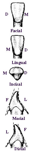
Figure 2.44. Mandibular Central Incisor
Figure 2-45. Mandibular Lateral Incisor |
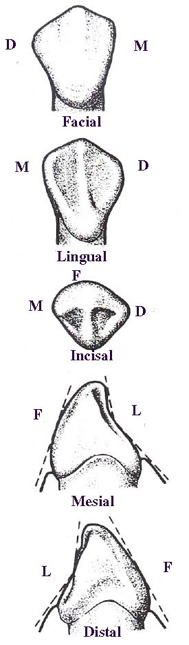
Figure 2-46. Madibular Cuspid |
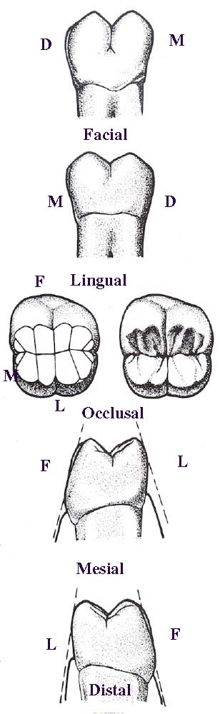
Figure 2-47. Mandibular First Bicuspid |
¨ Viewed on end, the incisal edge forms two curves that meet at the tip of the cusp. The mesial portion of the facial outline is convex, but the distal portion is slightly flattened. The mesial curve follows the alignment of the facial surfaces of the anterior teeth. The distal part of the facial outline conforms to the buccal surface alignment of posterior teeth. The cingulum appears uniformly curved on both sides.
¨ The mesial surface more nearly resembles that of the incisors than the mesial surface of the maxillary cuspid in outline. The facial outline is convex. The lingual outline is chiefly concave except near the cervical margin. The mesial surface is generally convex.
¨ The distal surface is shorter than the mesial surface but is of about the same width. The incisal portion is very convex both faciolingually and incisogingivally. The cervical portion is concave incisogingivally.
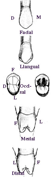
n Mandibular First Bicuspid
The mandibular first bicuspid (Figure 2-47) is the fourth tooth from the median line in the mandibular arch. It is the smallest and least typical of the bicuspids.
¨ The facial surface is shaped somewhat like a bell because the cervical portion is markedly constricted in comparison with the occlusal portion. The distal cusp ridge of the occlusal border is slightly longer than the mesial cusp ridge, and the distoincisal angle is more rounded than the mesioincisal angle. The distal portion of the surface is slightly shorter and broader than the mesial surface. The surface is convex.
¨ The lingual surface is much smaller than the facial surface because the lingual cusp is smaller than the facial cusp. The tip of the lingual cusp is closer to the mesial margin than to the distal margin. The surface is convex.
¨ The occlusal surface is marked by a strong facial cusp and a lingual cusp that may appear almost rudimentary. The marginal ridges are well defined. The strong lingual ridge of the facial cusp and the facial ridge of the lingual cusp may join, forming a transverse ridge. In this instance, the central groove would be very faint.
¨ The mesial surface is irregular in outline. From this aspect the tooth appears to be tipped lingually. The facial cusp forms most of the occlusal outline. The facial outline is very convex and the greatest convexity is in the cervical third. The lingual outline is fairly straight. Occlusocervically, the mesial surface is very convex in the occlusal portion and concave in the cervical portion.
¨ The distal surface is similar to the mesial surface.
n Mandibular Second Bicuspid
The mandibular second bicuspid (Figure 2-48) is the fifth tooth from the median line in the mandibular arch.
¨ The facial surface is very similar to the surface of the mandibular first bicuspid. The facial ridge is prominent, and the surface is convex.
¨ The lingual surface is similar to the surface of the mandibular first bicuspid, with the exception that there may be two cusps, the mesiolingual and the distolingual.
¨ The occlusal surface may appear in a number of forms. In the form pictured, the mesial and distal triangular fossae are quite distinct as they join the short central groove. There are three pits, the central, the mesial, and the distal.
¨ The mesial surface is similar to the surface of the mandibular first bicuspid, but it is more regular in outline. The surface is convex faciolingually. Occlusocervically, the occlusal portion is convex, and the cervical portion is concave.
¨ The distal surface is very similar to the mesial surface.
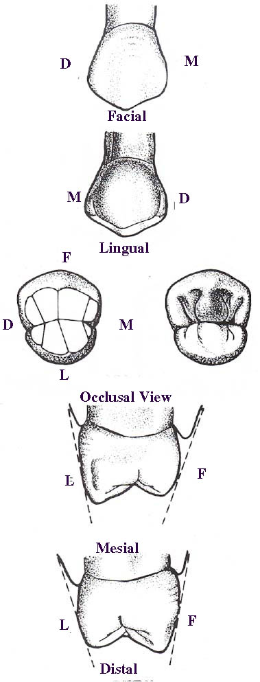
n Mandibular First Molar
The mandibular first molar (Figure 2-49) is the sixth tooth from the median line in the mandibular arch. It is also the largest tooth in the mandibular arch. The maxillary and mandibular first molars are often called "6-year" molars.
¨ The facial surface presents three cusps: the mesiofacial, the distofacial, and the distal. The mesiofacial cusp is the largest and the distal is the smallest. The distofacial cusp, though smaller than the mesiofacial cusp, may be slightly higher. The mesiofacial (facial) groove, which may end in a pit, separates the mesiofacial and distofacial cusps. The distofacial groove separates the distofacial and distal cusps. The facial surface is convex except at the grooves.
¨ The lingual surface has a mesiolingual cusp and a distolingual cusp, which are similar in outline. They are separated by the sharply defined lingual groove. The surface is slightly convex.
¨ The occlusal surface of this tooth, unlike the surface of the maxillary first molar, is formed by all five cusps and is trapezoidal in shape. There are three pits, the mesial,
 |
 |
 |
the central, and the distal. A central groove, which connects these pits, divides the occlusal surface into the lingual and facial halves. From the occlusal aspect, the mesiofacial cusp appears the largest and the distal cusp appears the smallest. ¨ The mesial surface is wider in the cervical portion than it is in the occlusal portion because the occlusal and middle thirds of the facial outline slope outward occlusocervically. The lingual outline is quite straight and nearly perpendicular. ¨ The distal surface is more symmetrical than the mesial surface because the facial outline is more nearly perpendicular than it is on the mesial surface. n Mandibular Second Molar The mandibular second molar (Figure 2-50) is the seventh tooth from the median line in the mandibular arch. It is one of the 12-years molars. ¨ The facial surface is almost symmetrical in outline, and the mesiofacial and distofacial cusps appear nearly equal in size. The two cusps are separated by the deep facial groove. There is no third cusp, as on the mandibular first molar. ¨ The lingual surface is symmetrical, but the mesiolingual cusp is slightly longer and bulkier than the distolingual cusp. The lingual groove is shorter and less distinct than the groove on the facial surface. |
¨ The occlusal surface is rectangular in shape. From this view the mesiofacial cusp appears slightly larger than the three other cusps. The occlusal surface has three pits, the mesial, the central, and the distal.
¨ The mesial surface resembles the mesial surface of the mandibular first molar; however, it is shorter. The facial outline is convex occlusocervically. The occlusal portion of the lingual outline is convex and the cervical portion is more nearly straight.
¨ The distal surface resembles the mesial surface.
Anatomy of Facial and Oral Structures
Below is a table showing the sequence of eruption of the primary dentition:


Below is a table showing the sequence of eruption of the permanent set:


Note: *b stands for buccal or toward the cheek; l stands for lingual or towards the tongue or palate.