 |
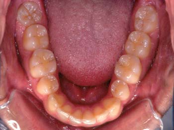 |
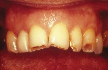
| Fig. 1 Purely physiological tooth surface loss | |
| Contents | Post-Test |
S. J. Davies1 R. J. M. Gray2 and A. J. E. Qualtrough3
This part of the series is devoted to tooth surface loss ( TSL ) not caused by caries or trauma. The management of this form of generalised TSL is included in this series because knowledge of occlusion is needed for both the diagnosis and, when indicated, treatment. There are, however, many other factors involved in the management of generalised TSL other than those associated with ‘occlusion’. These will also be discussed.
1*GDP, 73 Buxton Rd, High Lane, Stockport SK6 8DR; P/T Lecturer in Dental Practice, University Dental Hospital of Manchester, Higher Cambridge St.,Manchester M15 6FH;
2Honorary Fellow, University Dental Hospital of Manchester, Higher Cambridge St., Manchester M15 6FH
3Senior Lecturer and Honorary Consultantin Restorative Dentistry, University Dental Hospital of Manchester, Higher Cambridge St., Manchester M15 6FH
*Correspondence to : Stephen Davies, 73 Buxton Rd, High Lane, Stockport SK6 8DR
Email: stephen.j.davies@man.ac.uk
Tooth Surface Loss
Physiological?
Pathological?
Is the Tooth Surface loss Physiological or Pathological?
Tooth surface loss may be purely physiological (Fig. 1) and occurs as a natural consequence of ageing.1 Several factors, however, including erosion, abrasion and attrition can render tooth surface loss pathological (Fig. 2). As a result of this, symptoms may develop and treatment may be indicated. Although this chapter will deal with only pathological tooth surface loss, it is important to be able to recognise when tooth surface loss is purely physiological; it cannot be assumed that all tooth surface loss is pathological.
|
|
| Fig. 1 Purely physiological tooth surface loss | |
Refereed Paper © British Dental Journal 2002; 192: 11–23
|
Fig. 2 Pathological tooth surface loss may result in one or more of the following |
|
There appears to be no consensus as to what constitutes physiological tooth surface loss. It would be of assistance to practising dentists, if such criteria could be established. In the absence of accepted criteria of physiological tooth surface loss, those of pathological tooth surface loss are presented in Figure 2.
There are usually considered to be three reasons for non carious tooth surface loss. Abfractions should also be considered to be a cause of noncarious TSL.
Erosion is a chemical process in which the tooth surface is removed in the absence of plaque.2 Erosive factors may be either intrinsic or extrinsic. Extrinsic sources include drinks such as fresh fruit juices, carbonated drinks, cordials and alcoholic beverages; and some foods and industrial processes. Intrinsic sources include gastrooesophageal reflux and eating disorders, amongst others.
External agents which have an abrasive effect on the teeth include toothbrush bristles and dietary factors.
Attrition is a process in which tooth tissue is removed as a result of opposing tooth surfaces contacting during function or parafunction. Such direct contact occurs at proximal areas, on supporting cusps and on guiding surfaces during empty grinding movements.
It has been suggested that the stress lesion or abfraction is a consequence of eccentric forces on the natural dentition.3,4 The theory propounds tooth fatigue, flexure and deformation via biomechanical loading of the tooth structure, primarily at the cervical regions. Cusp flexure causes stress at the cervical fulcrum and results in loss of the overlying tooth structure. The lesion is typically wedge shaped with sharp line angles, but occlusal abfractions may present as circular invaginations. The magnitude of tooth tissue loss depends on the size, duration, direction, frequency and location of the forces. It should be remembered that abfractive lesions are caused by flexure and fatigue of susceptible teeth at sites that are usually distant from the point of loading. Other factors, such as erosion and abrasion may play a significant role in tooth tissue loss, but the initial force is the biomechanical loading.
Figure 3 shows a cervical cavity which is significantly subgingival and interestingly has caused a fenestration of the overlying attached gingiva. It is highly likely, because of the protection of the overlying soft tissue, that this lesion is an abfraction without any abrasive or erosive component.
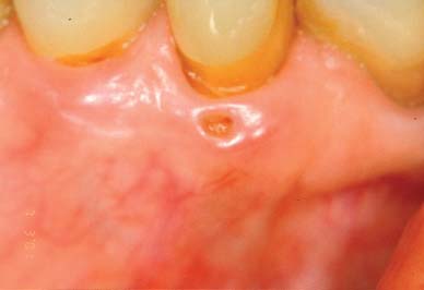 |
| Fig. 3 Abfraction |
Terminology:
Function
a normal movement
Parafunction
a normal movement at an abnormal frequency
Dysfunction
an abnormal movement
Bruxism is an important factor related to tooth surface loss. It is defined as the grinding of teeth during non functional movements of the masticatory system: it is a mandibular parafunction.
The wear is usually uniform when opposing teeth are affected. If bruxism is severe, either marked wear of occlusal surfaces will occur or, in cases of compromised periodontal support, tooth mobility may result. Bruxism can also be associated with muscle spasm, fractured teeth and restorations.5
Tooth surface loss or tooth wear cannot be taken as a sign that the patient is an active bruxist. Even if the cause of the TSL was bruxism the patient may no longer be bruxing. The signs of active bruxism are tongue scalloping and cheek ridging.6
Two aetiological models have been proposed:
There are, however, no reliable predictive indicators to suggest a simple relationship between the causes and effects of bruxism. It was once thought that ‘Type A personalities’ were more susceptible to stress related bruxism, but current teaching discounts this classification as a gross oversimplification and it is no longer generally used.
As proximal contacts wear in a normal dentition, there is a compensatory occlusal adjustment. This may happen naturally if the diet is abrasive ie this is as a result of function, not parafunction, and so should be not be considered pathological tooth surface loss caused by bruxism.
It has been found that the signs and symptoms of bruxism often disappear when occlusal therapy is aimed at the provision of an ideal occlusion: that is the careful elimination of interferences in the static and dynamic occlusion and the maximal distribution of occlusal load. Occlusal therapy should only be carried out after successful stabilisation splint usage, and careful ‘mock’ equilibration on accurately mounted study models. The need for a period of splint therapy is reinforced by the fact that strong clenching has the effect of compressing the periodontal ligament and rebound may take more than half an hour. This is of particular significance when the occlusion is either being assessed or equilibrated.
Furthermore, if the aim is to discourage a bruxist habit, this may be achieved by the patient’s intermittent use of an occlusal splint; a stabilisation splint or a localised occlusal interference splint, or in some cases a soft bite guard.
The patient for whom a diagnosis of pathological tooth surface loss is suspected, should be given a particular examination (Fig. 4). This is in addition to the normal examination of the articulatory system.
| Patient | Date | Age | |||
|
Normal | ||||
|
for a patient of this age? |
|||||
| Is there any evidence that it is progressive? | |||||
|
None |
|||||
|
EROSION | ||||
|
ABRASION |
|||||
|
ATTRITION |
|||||
|
|||||
| Known gastric reflux? | |||||
| Known parafunction? | |||||
| Other [specify]? | |||||
|
|||||
|
|||||
| Patient is worried about: | Appearance | ||||
| Tooth sensitivity | Tooth or restoration fracture | ||||
| Soft tissue comfort | Loosening teeth | ||||
|
|||||
| Centric Relation jaw registration | Models | ||||
| Photographs | Tooth measurements taken | ||||
| Ethyl chloride | |||||
This examination is designed to:
This question gives rise to Box 1 of the examination sheet (Fig. 4)
|
Normal | |
| Excessive | ||
| Extreme | ||
|
for a patient of this age? |
||
| Is there any evidence that it is progressive? | ||
| None | ||
| Weak | ||
| Strong | ||
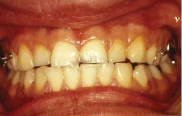
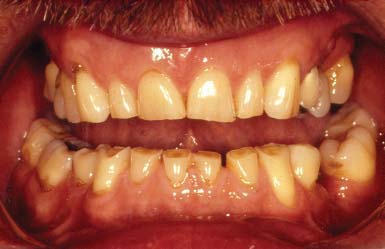
The features of pathological tooth surface loss: Pathological tooth surface loss may result in a change in the appearance of teeth, considered to be excessive with respect to the age of the patient (Fig. 5).7 There may be:
 |
 |
 |
| Fig. 5: Fig. 5a shows a young patient (18 years), who needs treatment, whereas in Fig. 5b a similar amount of TSL in an older (75 years) patient does not necessarily warrant intervention. The patient in Fig. 5c is 45 years old and his request for treatment is justified | ||
This question gives rise to Boxes 2, 3 of the examination sheet (Fig. 4).
|
EROSION | ||||
|
ABRASION |
|||||
|
ATTRITION |
|||||
|
|||||
| Known gastric reflux? | |||||
| Known parafunction? | |||||
| Other [specify]? |
The Features of Different Types of Tooth Surface Loss
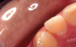
Despite the multi-factorial aetiology of tooth surface loss, certain clinical features may suggest a major contributory factor. Flattening of cusps or incisal edges and localised facets on occlusal or palatal surfaces would indicate a primarily attritional aetiology. Traditionally, cervical lesions caused purely by abrasion have sharply defined margins and a smooth, hard surface. The lesion may become more rounded and shallow if there is an element of erosion present. Once dentine is exposed, the clinical appearance is determined by the relative contribution of the aetiological factors. If wear is primarily attritional, then dentine tends to wear at the same rate as the surrounding enamel. Erosive lesions cause ‘cupping’ to form in the dentine. When erosion affects the palatal surfaces of upper maxillary teeth, there is often a central area of exposed dentine surrounded by a border of unaffected enamel.8
This question gives rise to Box 4 of the examination sheet (Fig. 4).
|
Physiological tooth surface loss is normal and results in a reduction in both vertical tooth height and horizontal tooth width.
In physiological tooth surface loss, vertical dimension is maintained by alveolar bone remodelling resulting in an elongation of the (dento-)alveolar process, similarly proximal wear is compensated by a constant forward pressure maintaining tooth to tooth contact.
If pathological vertical tooth surface loss has occurred then there is the possibility that a compensatory growth (‘dento-alveolar compensation’) may have occurred to some degree. This is an important consideration.
If tooth surface loss affecting the occlusal surfaces of the teeth has occurred, then one might expect to see a reduction in occlusal face height (vertical dimension of occlusion or VDO); or, expressed in a different way, an increase in the freeway space (FWS) could be anticipated. This may be further complicated by forward posturing of the mandible. It is often observed, however, that despite overall tooth surface loss, the freeway space and the resting facial height appear to remain unaltered primarily because of dento-alveolar compensation.
This is important with respect to patient assessment. If restoration of worn teeth is being planned then the extent of dento-alveolar compensation would appear to determine the dentist’s strategy; defining the need to carry out measures such as crown lengthening, to ensure the same VDO and FWS .
Nevertheless, the Fundamental Question is:
‘Does it matter if the patient’s VDO is increased during the restoration of the tooth surface loss (ie the FWS is reduced)?’
The answer is different for each patient. No occlusion can be said to be ‘wrong’ rather it is the case that in certain patients, at particular times in their lives, some occlusal patterns will not be tolerated. An occlusion can only be judged by the reaction of the tissues surrounding it, so it is with the issue of an increase in VDO.
In the case of a patient presenting with some dento-alveolar compensation, the clinician should assess whether or not that patient can tolerate an increased VDO (reduced FWS) by the construction of a stabilisation splint and/or provisional restorations.
Tooth Surface Loss
Has dento-alveolar compensation taken place?
The Incidence of Dento-Alveolar Compensation
It has been observed that in the normal adult dentition, the FWS remains constant and even in those patients who exhibit significant tooth surface loss the VDO is unaffected in 80% and a normal FWS of 3 mm is exhibited. If treatment of a patient within this group is necessary then crown lengthening procedures may be indicated. This will enable adequate reduction of the teeth at crown preparation so allowing the same VDO to be maintained. Alternatively restoration of the patient’s dentition may be provided at an increased VDO (reduced FWS). Some may argue that any increase in FWS should be proportionate to the degree of attrition.
This Question Gives Rise to Box 5 of the Examination Sheet (Fig. 4).
|
|||||
| Patient is worried about: | Appearance | ||||
| Tooth sensitivity | Tooth or restoration fracture | ||||
| Soft tissue comfort | Loosening teeth | ||||
Patient’s Wants and Needs
It is essential as in all areas of clinical practice to carefully consider the patient’s anxieties and desires in addition to the clinical features before advice is given.
Monitoring
Monitoring involves taking a series of repeated examinations and certain measurements over a period of time in order to assess whether a condition is progressive. Monitoring is essential in the management of tooth surface loss.
In the literature several methods of assessing tooth wear have been described including:
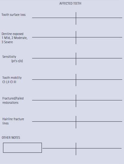
These are research tools and are not applicable to clinical practice. Therefore, a monitoring protocol to assess tooth surface loss is presented (Fig. 6a,b). It is easy to use and by this method it is easy to record the progression of tooth surface loss.
Fig. 6a Tooth surface loss monitoring
| Patient | Date | Age | |||||
| Primary aetiology |
EROSION |
||||||
|
ABRASION |
|||||||
|
ATTRITION |
|||||||
| RV no: | DATE OF LAST RV | INTERVAL | |||||
|
|
| Patient | Date |
|||
| OTHER SIGNS | ||||
| Active bruxism? | Tongue scalloping | |||
| Cheek ridging | ||||
| T.M.J. exam | Noise | |||
| Range of motion | ||||
| Tenderness to palpitation | ||||
| Muscle tenderness | ||||
| RECORDS TAKEN | ||||
| Photographs |
Impressions |
|||
| Measurements from A.D.J. | ||||
| ASSESSMENT at this time | ||||
| Non-progressive | ||||
| Maybe progressive [not marked and no symptoms] | ||||
| Progressive [obvious] | ||||
| Page 2 of 2 Progressive [obvious and symptomatic] | ||||
| TREATMENT at this time | ||||
Monitoring is, of course, only an option when baseline measurements have been taken. This emphasises the need for the dentist to examine and record tooth surface loss. To facilitate this a protocol for the initial examination of a patient with TSL has been presented (Fig. 4).
The key question to be answered is:
‘Does this Patient Need Treatment?’
There are no hard and fast rules and the need for treatment should be established after considering:
Monitoring (Figs 6a and 6)
This has already been discussed under the ‘Examination section’ and it represents a review process. Monitoring is the only way in which TSL can be assessed as being active or static. It is, therefore, the case that in most situations a period of monitoring should be carried out prior to considering active treatment.
Prevention
This is ‘treatment’ of future tooth surface loss. If the extent of existing tooth surface loss is considered to be acceptable, the appropriate treatment is clearly to try to prevent further TSL, which could render the patient needing restorative treatment. The form of the preventive treatment will be dependent on the aetiology of the TSL, so determining the cause is essential.
For a patient whose tooth surface loss is essentially caused by erosive fluids, advice regarding diet, the use of sugar free chewing gum, and the prescription of a fluoride mouthwash will almost certainly be indicated. It may be also necessary to liaise with the doctor if you suspect the patient suffers from a depressive illness.
If the wear is primarily caused by abrasion then examination and modification of the tooth cleaning habits will be indicated.
If the wear is caused by attrition, then the patient should be advised of any possible bruxist habits. The provision of one of three different sorts of splints could be considered. A soft bite guard can help in breaking a bruxist habit or simply will protect the teeth during the bruxist habit. A localised occlusal interference splint is designed to break the bruxist habit, and can be worn easily during the day. A stabilisation splint reduces bruxism by providing an ideal occlusion: it also enables the clinician to locate and record centric relation.9
Non-carious loss of tooth tissue may require treatment for one or more of the following reasons:
The latter may present a critical problem and both the need for restorative treatment and the complexity of that restorative treatment may depend upon whether or not dentoalveolar compensation has occurred, as previously discussed.
Where do We Start?
Careful planning regarding the reconstruction of a worn dentition is essential and, as in any other restorative treatment plan, the first decision to be made is whether or not the restorations will be designed to:
APPROACH The following criteria should be met if the conformative approach is to be considered:
If these criteria do not pertain, then a re-organised approach will need to be considered.
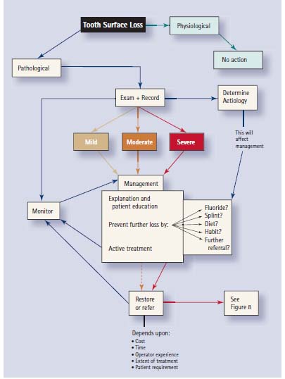
Whether or not a particular patient exhibiting TSL needs treatment or monitoring can only be determined by a complex series of decisions. An attempt to illustrate this process, relatively simply, is presented as Figure 7.
Fig. 7 Management of a patient with pathological tooth surface loss
|
|
The alternatives for treatment are equally complex if the decision has been taken that a patient needs treatment.
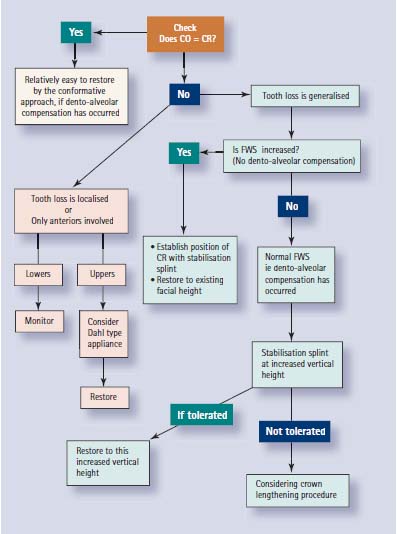
The process starts with an examination of the jaw relationship in which the patient is presently occluding (does CO occur in CR?).
From that decision it is possible to provide some structure to the treatment planning depending upon other factors. Figure 8 is an attempt to present this in a logical sequence.
|
|
Reprinted with permission.
British Dental Journal
Volume__, No.___, Date_______